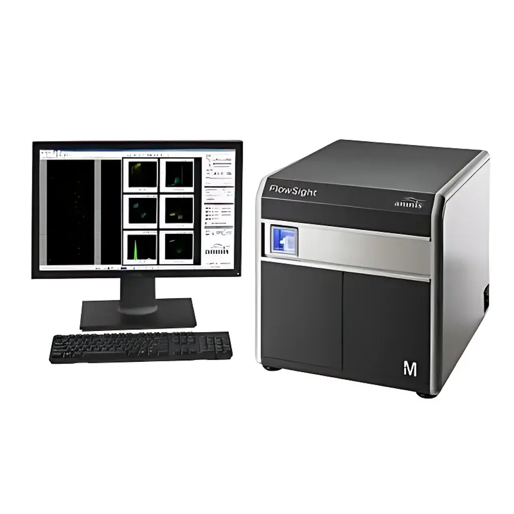
Merck Millipore FlowSight Quantitative Imaging Flow Cytometer
| Brand | Merck |
|---|---|
| Origin | USA |
| Manufacturer | Merck Millipore (acquired Amnis) |
| Product Type | Imported Instrument |
| Model | FlowSight |
| Pricing | Available Upon Request |
Overview
The Merck Millipore FlowSight Quantitative Imaging Flow Cytometer represents a paradigm shift in flow cytometry—bridging the gap between population-level statistics and single-cell morphological and functional resolution. Unlike conventional flow cytometers that rely solely on integrated fluorescence intensity and light scatter signals, the FlowSight integrates high-speed, quantitative digital imaging into the flow cytometric workflow. It employs time-delay integration (TDI) CCD imaging technology coupled with a multi-laser excitation platform to capture 12 simultaneous images per cell: one brightfield (BF), one darkfield (SSC), and up to ten fluorescence channels. This enables true image-based gating, morphometric analysis, subcellular localization quantification, and spatial fluorescence co-localization—all at throughput rates exceeding 1,000 cells/second. The system operates on principles of hydrodynamic focusing, laser-induced fluorescence, and synchronized TDI acquisition, delivering both statistical rigor and visual verification for complex biological questions in immunology, cell cycle dynamics, signaling translocation, and rare-event detection.
Key Features
- 12-channel imaging capability per cell: 1 brightfield, 1 side-scatter (SSC), and up to 10 fluorescence channels
- Multi-laser configuration: Standard 405 nm, 488 nm, 561 nm, and 642 nm lasers; optional 785 nm laser dedicated to enhanced SSC signal acquisition
- Sub-10 MESF (Molecules of Equivalent Soluble Fluorochrome) sensitivity—optimized for low-abundance antigen detection and weakly fluorescent probes
- Real-time autofocus and velocity-sensing optics ensure consistent image focus across variable flow rates and cell sizes
- Integrated 96-well autosampler option for unattended, reproducible high-throughput acquisition
- Proprietary TDI CCD detector with >10× higher signal-to-noise ratio than conventional PMT-based systems
- Image-based gating interface: Click any dot in a scatter plot or histogram to instantly retrieve its corresponding full-resolution cellular image
Sample Compatibility & Compliance
The FlowSight accepts standard suspension samples—including peripheral blood mononuclear cells (PBMCs), cultured adherent cells (after enzymatic detachment), primary tissue digests, and microorganisms—within typical flow cytometry size ranges (1–20 µm). Its fluidics architecture supports both sheath pressure–controlled and volumetrically calibrated acquisition modes, enabling absolute cell counting when paired with calibrated beads. The instrument complies with IEC 61010-1 safety standards for laboratory equipment and meets electromagnetic compatibility (EMC) requirements per EN 61326-1. Data integrity workflows support GLP/GMP-aligned environments through audit-trail–enabled software logging, user-access controls, and electronic signature compatibility. While not FDA-cleared as an IVD device, the FlowSight is widely used in preclinical research supporting regulatory submissions under ISO 13485 and 21 CFR Part 11–compliant data management practices.
Software & Data Management
IDEAS® software (v6.x or later) serves as the native acquisition, analysis, and reporting platform. It provides a unified environment for image processing, feature extraction (e.g., nuclear/cytoplasmic intensity ratios, object morphology, texture metrics), machine-learning–assisted classification (via built-in SVM and Random Forest tools), and batch analysis across thousands of acquired images. All raw image data are stored in standardized TIFF format with embedded metadata (laser power, filter set, gain, exposure time, sample ID), ensuring FAIR (Findable, Accessible, Interoperable, Reusable) data principles. IDEAS supports export to FCS 3.1 format for cross-platform compatibility with FlowJo™, Cytobank™, and other third-party analysis suites. Audit trails record all gating decisions, parameter adjustments, and export events—critical for traceability in regulated research settings.
Applications
- Cell cycle & mitotic staging: Discrimination of prophase, metaphase, anaphase, and telophase via chromatin condensation and spindle morphology—beyond standard DNA content analysis
- Multiparametric immunophenotyping: Simultaneous identification of ≥8 immune subsets (e.g., CD3⁺/CD4⁺ T cells, CD14⁺ monocytes, CD123⁺ pDCs) with spatial confirmation of marker co-expression and internalization
- Transcription factor translocation: Quantitative measurement of NF-κB nuclear import kinetics using DAPI/FITC dual-channel image correlation and pixel-wise nuclear masking
- Phagocytosis & pathogen uptake: Direct visualization and quantification of internalized particles or microbes within phagosomes using differential labeling and z-stack–like optical sectioning
- Apoptosis & membrane integrity assessment: Morphological validation of Annexin V/PI staining patterns alongside blebbing, shrinkage, and nuclear fragmentation features
FAQ
What distinguishes FlowSight from spectral or conventional flow cytometers?
The FlowSight captures actual digital images—not just intensity values—enabling morphological validation, spatial fluorescence analysis, and image-based gating. Spectral cytometers resolve fluorochrome spectra but do not provide spatial context.
Can FlowSight perform absolute cell counting?
Yes—when used with NIST-traceable calibration beads and volumetric acquisition mode, it delivers accurate absolute counts per microliter without requiring secondary reference standards.
Is IDEAS software compatible with modern macOS or Linux systems?
IDEAS runs natively on Windows 10/11 (64-bit). Virtualized deployment on macOS or Linux is possible via supported hypervisors, though hardware-accelerated image rendering is optimized for Windows.
Does FlowSight support live-cell imaging or time-lapse analysis?
No—it is designed for fixed or stabilized suspension samples. The high-speed imaging process requires stable hydrodynamic conditions incompatible with long-term viability maintenance.
How is data storage managed for large imaging datasets?
Raw image files are stored in compressed TIFF format; IDEAS includes lossless compression options and hierarchical data organization (experiment → sample → acquisition → event). Typical 100,000-cell experiments occupy ~2–4 GB depending on channel count and bit depth.
Related Products


