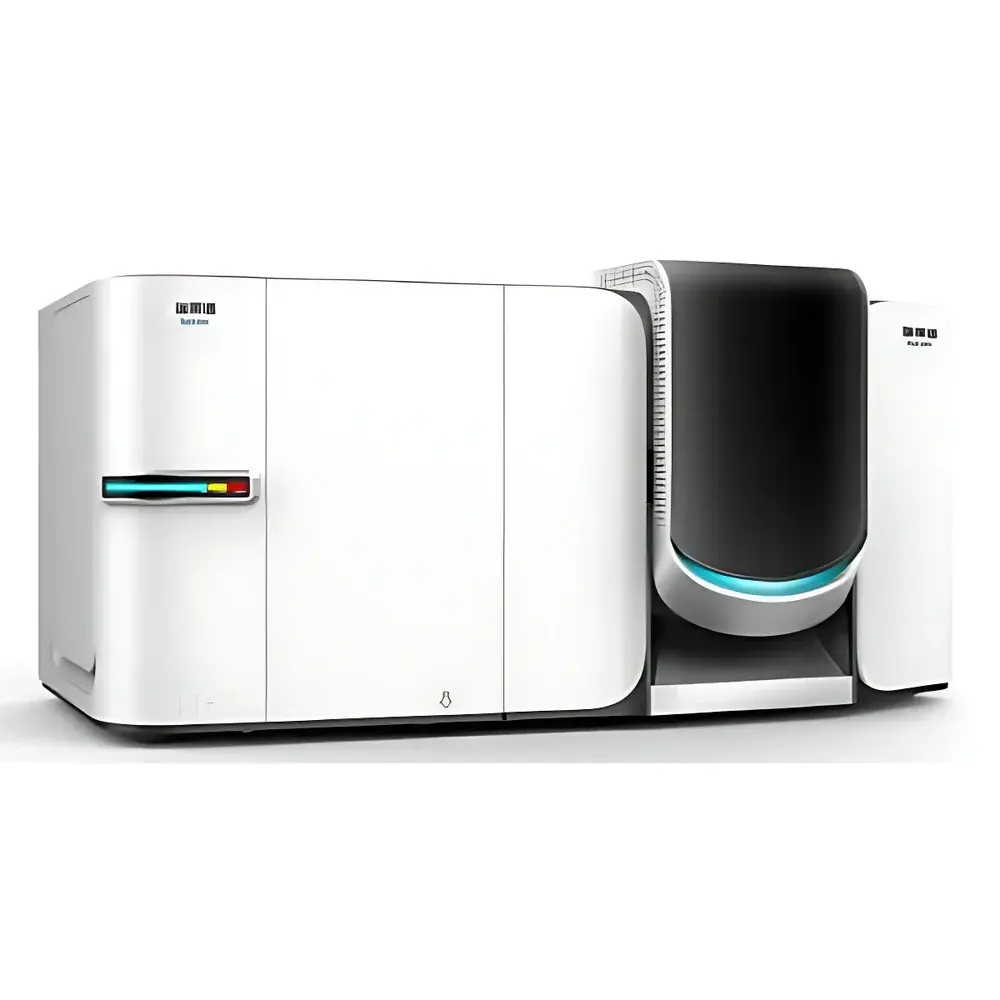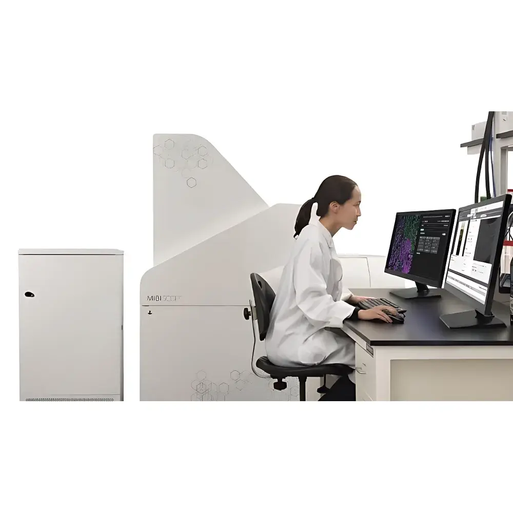
MIBIscope Ultra-High-Resolution Tissue Mass Spectrometry Imaging System
| Origin | USA |
|---|---|
| Manufacturer Type | Authorized Distributor |
| Origin Category | Imported |
| Model | MIBIscope |
| Instrument Type | Time-of-Flight (TOF) Mass Spectrometer |
| Application Domain | Universal |
| Spatial Resolution | 350 nm – 1 µm |
| Sensitivity Ratio | >10:1 |
| Stability | Excellent |
| Data Acquisition Rate | 50 Gbit/s |
| Mass Accuracy | <80 ppm |
Overview
The MIBIscope Ultra-High-Resolution Tissue Mass Spectrometry Imaging System is a purpose-built TOF-based imaging platform engineered for spatially resolved, multiplexed protein detection in intact tissue sections. Leveraging Multiplexed Ion Beam Imaging (MIBI) technology, it combines secondary ion mass spectrometry (SIMS) with high-brightness, isotopically pure metal-tagged antibodies to enable simultaneous quantification of over 40 protein biomarkers at subcellular spatial resolution. Unlike conventional immunofluorescence or MALDI-based approaches, MIBIscope operates on a deterministic ion beam rastering principle—employing a focused, multi-element primary ion beam (e.g., O−, Cs+) to sputter and ionize labeled epitopes with minimal lateral diffusion. This architecture ensures quantitative fidelity across the full dynamic range (>5 logs), preserves native tissue architecture, and eliminates spectral overlap inherent in optical multiplexing. The system is designed for integration into clinical research laboratories and translational pathology workflows where reproducible, spatially contextualized proteomic mapping is required for tumor microenvironment characterization, immune cell phenotyping, and biomarker co-localization studies.
Key Features
- Submicron spatial resolution (350 nm minimum pixel size) enabled by precision ion optics and high-stability stage positioning
- Simultaneous detection of ≥40 metal-conjugated antibodies using isotopically resolved TOF mass analysis
- Quantitative dynamic range exceeding 105, validated against NIST-traceable standards
- Integrated optical and scanning electron detector (SED) imaging for real-time ROI selection and registration
- Automated workflow from slide loading to TIFF export—fully compatible with standard FFPE and frozen tissue preparation protocols
- Robust vacuum architecture with differential pumping stages ensuring stable beam current and signal reproducibility across multi-hour acquisitions
- Compliance-ready data handling supporting audit trails, user access controls, and electronic signatures per FDA 21 CFR Part 11 requirements
Sample Compatibility & Compliance
MIBIscope accepts standard glass-mounted tissue sections (4–10 µm thickness), including formalin-fixed paraffin-embedded (FFPE), frozen, and decalcified bone specimens. Antibody conjugation follows standardized metal-tagging protocols (e.g., Maxpar® reagents), with validation kits available for common immune-oncology markers (CD3, CD4, CD8, FOXP3, PD-1, etc.). The platform meets ISO/IEC 17025:2017 criteria for analytical instrument qualification and supports GLP-compliant study execution. All acquisition metadata—including dwell time, beam current, mass calibration parameters, and stage coordinates—is embedded in TIFF headers and archived alongside raw .imzML files for regulatory traceability.
Software & Data Management
Acquisition and analysis are managed through MIBI Analyst™ v4.x, a validated desktop application built on a modular C++/Python framework. The software provides real-time spectral preview, automated mass drift correction, and pixel-wise isotope ratio normalization. Raw data is stored in open-format .imzML (Imaging Mass Spectrometry Markup Language) compliant with MSI Consortium specifications. Quantitative outputs are exported as calibrated TIFF stacks (16-bit or 32-bit float), compatible with downstream tools including HALO™, Visium Spatial Gene Expression Suite, and custom Python/R pipelines. Audit logs record all user actions, parameter modifications, and file exports—enabling full compliance with 21 CFR Part 11 and EU Annex 11 requirements for electronic records in regulated environments.
Applications
- Spatially resolved immune profiling in tumor microenvironments (TME)
- Multi-parametric cell segmentation and phenotyping without signal crosstalk
- Validation of novel therapeutic targets via co-expression analysis across tissue compartments
- Longitudinal biomarker monitoring in preclinical xenograft and PDX models
- Correlative imaging with H&E, IHC, and digital pathology platforms
- Development of spatially informed diagnostic algorithms for companion diagnostics
FAQ
What sample preparation protocols are validated for MIBIscope?
Standard FFPE deparaffinization, antigen retrieval (heat-induced or enzymatic), and metal-tagged antibody incubation per Maxpar® guidelines. Frozen sections require acetone fixation and no antigen retrieval.
Is mass calibration performed automatically during acquisition?
Yes—internal reference ions (e.g., 115In+, 165Ho+) are introduced continuously via a dedicated calibration gas inlet, enabling real-time mass axis correction with <±5 ppm deviation over 8-hour runs.
Can MIBIscope data be integrated with transcriptomic spatial datasets?
Absolutely—TIFF outputs retain native pixel geometry and coordinate metadata, enabling direct registration with Visium, Xenium, or MERFISH datasets using elastix or BigWarp alignment tools.
What level of IT infrastructure is required for data storage?
A single 350-nm scan (800×800 µm², 40 channels) generates ~250 GB of raw .imzML data; we recommend NAS storage with ≥10 GbE connectivity and RAID 6 redundancy for production labs.
Does the system support Good Manufacturing Practice (GMP) environments?
While primarily deployed in R&D and clinical trial support labs, MIBIscope’s software architecture, electronic logbooks, and IQ/OQ/PQ documentation packages are fully extensible to GMP-compliant manufacturing of companion diagnostics.