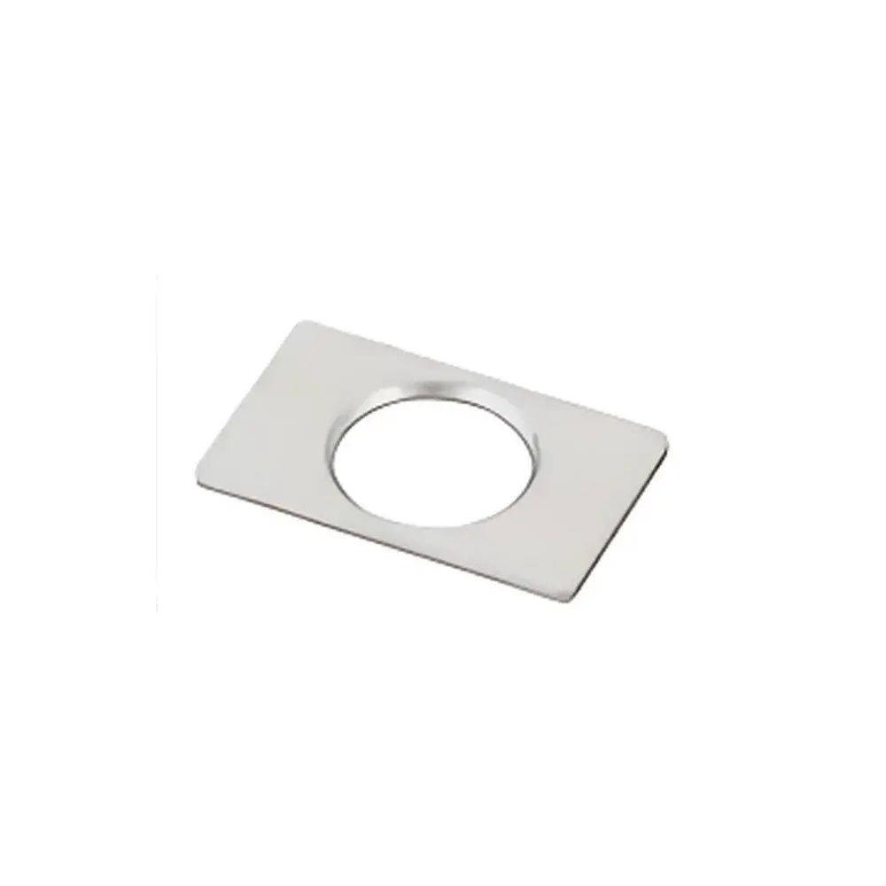
NARISHIGE CP-2 Chamber Plate
| Brand | NARISHIGE |
|---|---|
| Origin | Japan |
| Model | CP-2 |
| Material | Anodized Aluminum with Electrical Insulation |
| Observation Port Diameter | Ø9 mm |
| Dimensions (W × D × T) | 12 mm × 19 mm × 1 mm |
| Weight | ~0.4 g |
| Compliance | Designed for chronic in vivo rodent neurophysiology applications |
Overview
The NARISHIGE CP-2 Chamber Plate is a precision-engineered, ultra-lightweight mounting interface designed specifically for chronic cranial window and intracranial electrode implantation studies in murine models. Constructed from high-purity aluminum and subjected to a controlled anodization process followed by electrical insulation coating, the CP-2 provides mechanical stability, electrochemical inertness, and dielectric integrity—critical requirements when interfacing with sensitive electrophysiological recording systems or optical imaging setups. Its measurement principle is purely mechanical and geometric: the plate serves as a rigid, dimensionally stable reference platform that integrates seamlessly with NARISHIGE’s stereotactic frame systems (e.g., SR-6M, MO-10) to enable repeatable, sub-millimeter repositioning of microdrives, micromanipulators, or optical fibers across longitudinal experimental sessions. Unlike disposable polymer-based adapters, the CP-2 is reusable, autoclavable (per manufacturer guidelines), and compatible with standard craniotomy protocols involving dental acrylic anchoring.
Key Features
- Ultra-low mass (~0.4 g) minimizes mechanical load on the skull and reduces motion artifact during awake-behaving or head-fixed recordings
- Ø9 mm centered observation port enables unobstructed optical access for two-photon microscopy, epifluorescence, or fiber photometry without requiring lens repositioning
- Anodized aluminum substrate with certified electrical insulation layer ensures >1012 Ω surface resistivity—preventing signal leakage or ground-loop interference in extracellular electrophysiology
- 1 mm uniform thickness and precisely machined 12 mm × 19 mm footprint guarantee flatness tolerance < ±2 µm, essential for accurate stereotactic coordinate registration
- Chemically resistant surface withstands repeated exposure to ethanol, hydrogen peroxide, and common surgical disinfectants without degradation
- Compatible with NARISHIGE’s CP-series accessory ecosystem including CP-1 (Ø5 mm port), CP-10 (Ø10 mm countersunk port), and CF-10 frame clamps
Sample Compatibility & Compliance
The CP-2 is validated for use in C57BL/6, BALB/c, and CD-1 mice (typically 20–35 g body weight) undergoing chronic cranial window implantation, tetrode array insertion, or GRIN lens stabilization. It conforms to ISO 10993-5 (biological evaluation of medical devices — cytotoxicity testing) for material safety in prolonged tissue contact applications. While not a medical device per FDA classification, its design adheres to GLP-aligned documentation practices for preclinical neuroscience instrumentation. The anodized aluminum composition meets RoHS Directive 2011/65/EU restrictions on hazardous substances. No animal-derived components are used in manufacturing.
Software & Data Management
The CP-2 operates independently of software—it is a passive mechanical component. However, its dimensional repeatability directly supports digital workflow integration: coordinates registered during initial surgery (e.g., bregma-to-plate-center offset) can be imported into stereotactic planning software such as Stereotaxic Atlas Tools (SAT), Paxinos & Franklin’s Mouse Brain Atlas modules, or custom MATLAB/Python scripts for multi-session trajectory alignment. When used with NARISHIGE’s digital micromanipulators (e.g., MN-403), the CP-2 enables sub-5 µm positional reproducibility across days—facilitating longitudinal analysis of neural circuit dynamics under controlled experimental conditions.
Applications
- Chronic two-photon calcium imaging through thinned-skull or glass-window preparations
- Longitudinal single-unit and local field potential (LFP) recordings using movable microdrives
- Fiber photometry and optogenetic stimulation with fixed-depth optical fiber implants
- Combined electrophysiology-optics experiments requiring simultaneous electrical shielding and optical transparency
- Development and validation of closed-loop behavioral paradigms in head-fixed mice
- Standardized cranial mounting for multi-lab collaborative studies requiring inter-subject coordinate normalization
FAQ
Can the CP-2 be sterilized between uses?
Yes—autoclaving at 121°C, 15 psi for 20 minutes is supported; however, repeated cycles (>10) may gradually reduce surface insulation integrity. Ethanol wipe-down and UV irradiation are recommended for routine decontamination.
Is adhesive required for skull attachment?
Yes—dental acrylic (e.g., C&B Metabond or Super-Bond C&B) is the standard bonding agent. Cyanoacrylate adhesives are not recommended due to exothermic polymerization and potential neurotoxicity.
How does the CP-2 differ from the CP-1 and CP-10 variants?
The CP-1 features a smaller Ø5 mm port optimized for single-fiber or microelectrode access; the CP-10 offers a Ø10 mm countersunk port for larger-diameter optics or multi-channel probes; the CP-2 balances optical access and structural rigidity with its Ø9 mm non-countersunk port.
Does the CP-2 interfere with MRI or CT imaging?
No—aluminum generates negligible susceptibility artifacts in 7T and lower MRI systems; it is radiolucent in micro-CT, enabling co-registration of surgical landmarks with post-implant imaging data.


