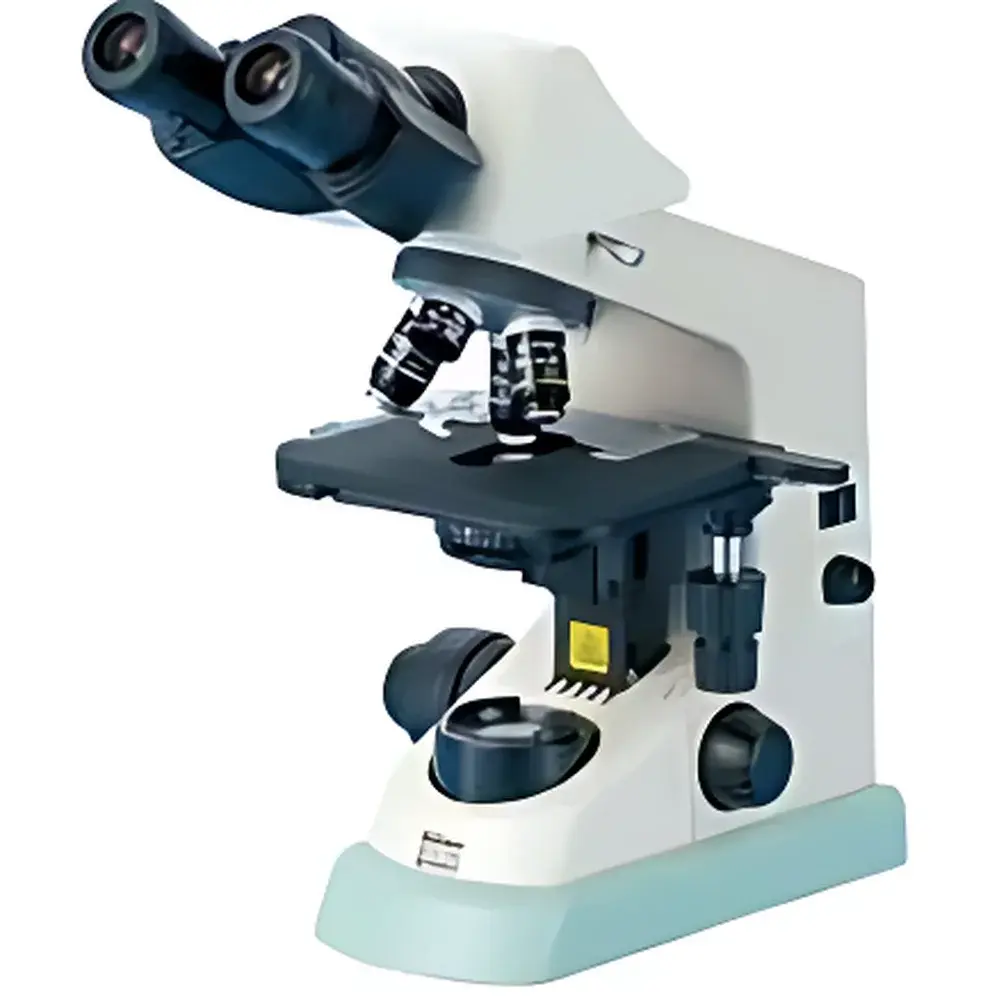
NIKON Eclipse E100 Biological Upright Microscope with Trinocular Head and LED Illumination
| Brand | NIKON |
|---|---|
| Origin | Japan |
| Model | E100 Trinocular |
| Optical System | CFI Infinity-Corrected |
| Observation Head | E2-TF Trinocular, 30° Inclination, Interpupillary Distance 47–75 mm |
| Eyepieces | CFI E 10×/18 (standard), optional CFI E 15×/12 |
| Objective Turret | Quadruple Nosepiece |
| Objectives | CFI BE Plan Achromat 4×, 10×, 40×, 100× (oil), optional 20×, 60× |
| Condenser | Abbe Condenser, NA 1.25, 33 mm Filter Holder |
| Focusing | Coaxial Coarse/Fine Focus, Fine Focus Graduation 2 µm, Coarse Travel 37.7 mm/rev, Fine Travel 0.2 mm/rev, Total Travel 22 mm |
| Stage | Double-Layer Mechanical Stage, 155 × 134 mm |
| Illumination | High-Efficiency White LED (60,000 h lifetime), Constant CCT across intensity range, IR-free, low heat, low power consumption |
| Accessories | Phase Contrast Annuli (10×, 40×, 100×), Darkfield Rings (10×, 20×, 40×), Pointer, Mirror, Power Cord Clip, Protective Packaging (Aluminum Case) |
| Anti-Fungal Treatment | Applied to eyepieces, objectives, and observation tube |
Overview
The NIKON Eclipse E100 is a high-performance upright biological microscope engineered for routine teaching laboratories, quality control environments, and entry-level research applications requiring reliable, reproducible optical imaging. Built upon Nikon’s proprietary CFI (Chromatic and Field Flattened Infinity) optical architecture, the E100 delivers corrected plan-achromatic imaging across the full field of view—minimizing field curvature and lateral chromatic aberration while maintaining extended working distances and improved numerical aperture performance. Its upright configuration supports standard glass slide-based specimen observation under brightfield, phase contrast, and darkfield modalities. The system integrates an advanced white LED illumination module that eliminates infrared emission, reduces thermal load on live or temperature-sensitive samples, and ensures stable color temperature (CCT) across the full dimming range—a critical advantage over traditional halogen systems where spectral shift compromises quantitative image consistency.
Key Features
- CFI BE Plan Achromat Objectives: Specifically designed for the E100 platform, these anti-fungal coated objectives deliver flat, high-contrast images at 4×, 10×, 40×, and 100× (oil immersion), with optional 20× and 60× magnifications available for expanded resolution flexibility.
- Trinocular Observation Head (E2-TF): 30° inclined, Siedentopf-style head with adjustable interpupillary distance (47–75 mm) and ±5 diopter eyepiece compensation; enables simultaneous visual observation and digital imaging via C-mount or dedicated microscope camera adapters.
- LED Illumination System: 60,000-hour rated lifetime, energy-efficient (≤5 W typical operation), zero IR emission, and thermally stable output—eliminating bulb replacement cycles and reducing long-term operational cost and downtime.
- Ergonomic Coaxial Focus Mechanism: Dual-speed coaxial coarse/fine focus with torque-adjustable coarse knob; fine focus graduation of 2 µm per division supports precise Z-axis positioning required for multiplane imaging and morphological documentation.
- Abbe Condenser (NA 1.25): Integrated position-indexed aperture diaphragm and objective-specific alignment markers (for 4×, 10×, 40×, 60×, 100×) ensure rapid, repeatable Köhler illumination setup without manual recalibration.
- Anti-Fungal Protection: Proprietary anti-mold coating applied to ocular lenses, objective barrels, and internal optical pathways—validated for sustained operation in high-humidity laboratory environments per ISO 8597:2017 environmental classification guidelines.
Sample Compatibility & Compliance
The E100 accommodates standard 1″ × 3″ (25 × 76 mm) glass slides and 24 × 50 mm coverslips. Its mechanical stage (155 × 134 mm) provides ample travel for multi-well plate scanning and tissue section mapping. Optional phase contrast and darkfield accessories support unstained live-cell observation and subcellular granule visualization without fixation or staining—enabling compliance with GLP-aligned cytology workflows. While not certified for clinical diagnostic use per FDA 21 CFR Part 866, the instrument meets IEC 61000-6-3 (EMC emissions) and IEC 61010-1 (safety for laboratory equipment) standards. Its LED light source conforms to IEC/TR 62778 for photobiological safety (RG0 classification), eliminating retinal hazard concerns during prolonged operator use.
Software & Data Management
The trinocular port is compatible with third-party USB 3.0 and HDMI-enabled digital cameras (e.g., AmScope MU series, OMAX 10MP models) and supports integration with open-source acquisition platforms such as Micro-Manager v2.x and commercial solutions including NIS-Elements BR (via optional adapter). Image metadata—including objective ID, magnification, illumination intensity, and timestamp—is embedded in TIFF/OME-TIFF export formats. Audit trail functionality is maintained when paired with validated LIMS or ELN systems compliant with 21 CFR Part 11 requirements—provided user authentication, electronic signatures, and change history logging are implemented at the software layer.
Applications
- Routine histopathology education and undergraduate biology labs
- Microbial morphology assessment (bacteria, yeast, algae) in QC microbiology
- Live-cell phase contrast imaging of mammalian and insect cell cultures
- Plant tissue section analysis and pollen grain identification
- Basic hematology screening (blood smears, parasite detection)
- Material inspection of thin polymer films and crystalline structures
FAQ
Is the E100 suitable for fluorescence imaging?
No—the E100 lacks a fluorescence excitation light path, dichroic mirrors, or emission filter slots. It is optimized for transmitted-light techniques only.
Can the LED illumination intensity be controlled externally?
Yes—intensity adjustment is performed via a front-panel rotary dial with 0–100% linear response; no external controller is required, though analog 0–10 V input compatibility is available via optional accessory interface.
What is the maximum usable magnification with the standard 10× eyepiece?
With the 100× oil immersion objective and 10× eyepiece, total magnification reaches 1000×—within the practical resolution limit (~0.2 µm) of the CFI BE optical design under optimal Köhler illumination.
Does the E100 support motorized focusing or stage automation?
No—motorization is not natively supported. The E100 is a manually operated platform intended for cost-sensitive, high-reliability educational and industrial QA settings.
Are replacement parts and service manuals available internationally?
Yes—NIKON-certified service centers in EMEA, APAC, and North America provide authorized calibration, repair, and genuine spare optics (objectives, condensers, LED modules) with traceable certification per ISO/IEC 17025.


