NIKON Patch-Clamp Electrophysiology System
| Brand | NIKON |
|---|---|
| Origin | USA |
| Manufacturer Type | Authorized Distributor |
| Import Status | Imported |
| Model | Patch-Clamp Electrophysiology System |
| Pricing | Available Upon Request |
Overview
The NIKON Patch-Clamp Electrophysiology System is a fully integrated, high-fidelity platform engineered for single-channel and whole-cell voltage-clamp and current-clamp recordings in live cells. Based on the fundamental principles of giga-seal patch-clamp electrophysiology—first established by Neher and Sakmann—the system enables precise measurement of picoampere (pA)-scale ionic currents across nanoscale membrane patches. The technique relies on forming a high-resistance (>1 GΩ) seal between a fire-polished glass micropipette and a cellular membrane, isolating a microscopic patch (typically 1–10 µm²) containing one or a few ion channels. By applying controlled voltage steps or ramp protocols via a feedback-controlled amplifier, the system resolves stochastic gating behavior—including open probability, mean open/closed times, conductance states, and ligand- or voltage-dependent kinetics—with sub-millisecond temporal resolution. Designed for rigorous physiological experimentation, the platform supports excised (inside-out, outside-out), cell-attached, whole-cell, and perforated-patch configurations, enabling systematic interrogation of channel pharmacology, modulation by intracellular second messengers, and structure-function relationships under defined ionic and biochemical environments.
Key Features
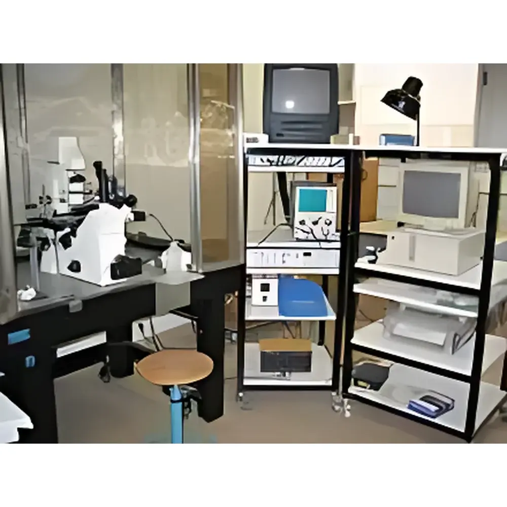
- Modular architecture integrating NIKON Ti-series inverted fluorescence microscope with motorized Z-stage, high-NA objectives (40×/60× water immersion), and LED-based epifluorescence illumination for simultaneous optical monitoring and electrophysiological recording
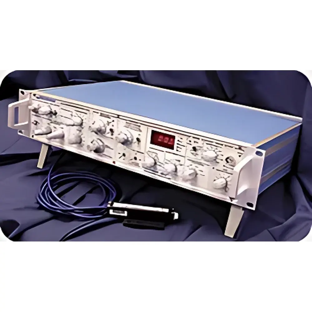
- High-stability Axopatch 200B or 700B headstage amplifiers with low-noise analog circuitry, capacitive compensation, and adjustable series resistance (Rs) correction for accurate whole-cell voltage control
- 8-channel computer-controlled perfusion system (PS25-Cell optional) with independent temperature regulation (20–42 °C, ±0.1 °C stability) and rapid solution exchange (<100 ms)
- SECM-compatible micromanipulator (Sutter MP-285 or equivalent) with sub-micron positioning resolution and vibration-damped kinematic coupling to the microscope stage
- Precision pipette puller (Sutter P-1000 or equivalent) for reproducible fabrication of borosilicate or quartz electrodes with tip resistances ranging from 2–10 MΩ
- Active anti-vibration optical table with grounded Faraday cage enclosure to minimize mechanical drift and electromagnetic interference during low-current measurements
- Dual-monitor workstation with dedicated data acquisition hardware (e.g., Digidata 1550B) and real-time digitization at up to 500 kHz sampling rate
Sample Compatibility & Compliance
The system supports primary neurons, cardiomyocytes, HEK293T, CHO, and other adherent or suspension mammalian cells; oocytes; and small tissue slices (≤300 µm thickness). All hardware and software components comply with ISO 13485 design controls for research-use-only (RUO) instrumentation. Data acquisition workflows adhere to GLP-compliant metadata tagging (time stamp, electrode ID, solution composition, holding potential), and optional audit-trail modules support 21 CFR Part 11 readiness for preclinical pharmacology studies. Electrical safety conforms to IEC 61010-1; optical components meet ANSI Z80.10 standards for laser safety classification where applicable.
Software & Data Management
Acquisition and analysis are performed using Clampex (Molecular Devices) or PatchMaster (HEKA) software suites, both supporting protocol-driven experiment automation, online leak subtraction, capacitance compensation, and batch-mode analysis of dwell-time histograms, amplitude distributions, and conductance variance. Raw data are stored in standardized ABF or HDF5 formats with embedded experimental parameters. Integrated export pipelines enable direct transfer to MATLAB, Python (via Neo or Axograph libraries), or commercial statistical platforms (GraphPad Prism, SAS) for kinetic modeling (e.g., hidden Markov modeling, maximum likelihood fitting). Metadata schema includes full traceability of pipette calibration, bath solution lot numbers, and environmental conditions (temperature, humidity, CO₂).
Applications
- Characterization of voltage-gated Na⁺, K⁺, Ca²⁺, and ligand-gated (e.g., nAChR, GABAₐ, NMDA) channel biophysics under physiological and pathophysiological conditions
- Cardiovascular pharmacology: assessment of drug-induced hERG blockade, late Na⁺ current enhancement, or calcium handling abnormalities in human iPSC-derived cardiomyocytes
- Mechanistic studies of channelopathies (e.g., Long QT syndrome, epilepsy-associated KCNQ2 mutations) using CRISPR-edited isogenic cell lines
- Real-time monitoring of intracellular signaling cascades via co-recorded Ca²⁺ fluorescence (e.g., GCaMP) and membrane current
- High-content functional screening of compound libraries against recombinant ion channel targets expressed in stable cell lines
- Investigation of synaptic transmission dynamics in acute brain slices using dual whole-cell recordings
FAQ
What is the minimum detectable current resolution of this system?
The Axopatch 200B/700B amplifier achieves RMS noise ≤50 fA (0.1–10 kHz bandwidth) in low-noise mode, enabling reliable detection of single-channel events ≥0.1 pA.
Can the system perform simultaneous fluorescence imaging and electrophysiology?
Yes—the NIKON Ti platform supports epifluorescence, TIRF, and confocal-ready configurations with optically isolated light paths and synchronized trigger outputs for frame-locked acquisition.
Is temperature control available for both bath and perfusate solutions?
The PS25-Cell perfusion module provides independent heating/cooling of all eight channels, while the microscope stage heater maintains bath temperature within ±0.1 °C of setpoint.
Does the software support automated patch formation algorithms?
While fully automated gigaseal formation is not standard, Clampex and PatchMaster offer programmable pressure/voltage protocols and real-time impedance monitoring to assist in seal optimization.
Are validation documents (IQ/OQ) available for regulated environments?
Yes—customizable installation and operational qualification protocols are provided upon request, aligned with ASTM E2500 and EU Annex 11 guidance for instrument lifecycle management.
Related Products


