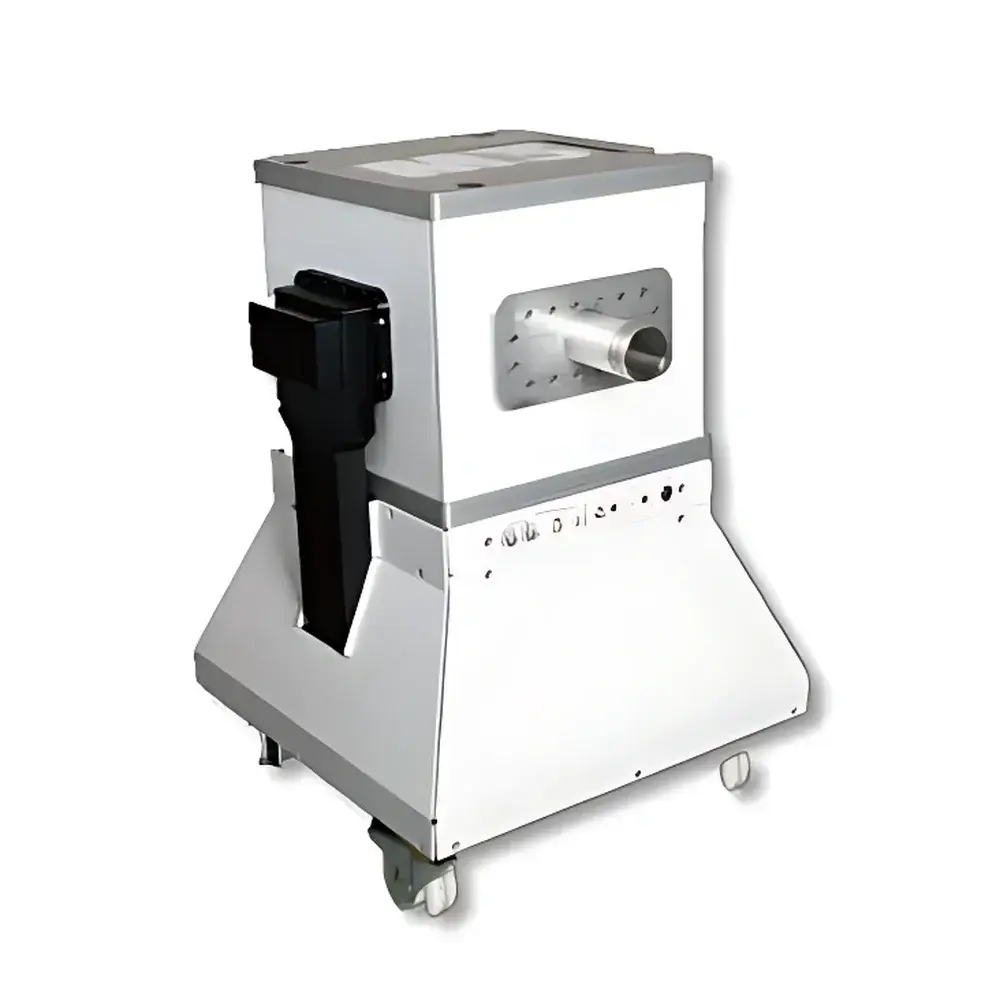
NIUMAG Aspect M3-mice Small Animal MRI System
| Brand | NIUMAG |
|---|---|
| Origin | Israel |
| Manufacturer Type | Authorized Distributor |
| Origin Category | Imported |
| Model | M3-mice |
| Instrument Type | Preclinical Magnetic Resonance Imaging (MRI) System |
| Magnet Type | Permanent Magnet, 1.0 Tesla |
| Spatial Resolution | ≤ 235 µm (isotropic, typical in vivo) |
| Scan Time | As low as 8–9 minutes per sequence (T1- or T2-weighted) |
| Cooling | Passive (no cryogens or chillers required) |
| Shielding | Self-shielded, zero fringe field beyond system footprint |
| Compliance | Designed for ISO 13485-aligned manufacturing and GLP-compliant preclinical research environments |
| Software Platform | Aspect ImageSuite™ v5.x (FDA 21 CFR Part 11–ready audit trail, DICOM 3.0 export, multi-sequence reconstruction pipeline) |
Overview
The NIUMAG Aspect M3-mice is a compact, self-shielded, 1.0 Tesla permanent magnet preclinical MRI system engineered exclusively for high-fidelity in vivo imaging of mice. Unlike superconducting systems requiring liquid helium and RF-shielded rooms, the M3-mice employs a passively cooled, high-stability permanent magnet architecture—eliminating cryogenic infrastructure, minimizing facility footprint, and enabling deployment directly behind biosafety level 2 (BSL-2) containment barriers. Its magnetic field homogeneity ( 1000 T/m/s) support robust acquisition of T1-, T2-, and proton density–weighted images with sub-250 µm isotropic resolution under standard in vivo conditions. The system operates on standard 230 V/50 Hz power without dedicated HVAC or RF shielding, making it suitable for core imaging facilities, pharmacology labs, and longitudinal phenotyping units where space, operational cost, and regulatory traceability are critical constraints.
Key Features
- Compact 1.0 T permanent magnet design: No cryogens, no quench risk, zero external fringe field—enables installation in standard laboratory spaces including BSL-2 animal housing corridors.
- Integrated gradient coil and RF probe optimized for murine anatomy: Dual-tuned (¹H/¹⁹F) volume coil included; optional surface coils available for region-specific SNR enhancement.
- Full-featured pulse sequence library: Includes spin echo (SE), fast spin echo (FSE), gradient echo (GRE), inversion recovery (IR), and diffusion-weighted imaging (DWI) sequences—preconfigured for murine brain, abdomen, heart, and musculoskeletal applications.
- Automated shimming and B₀ mapping: Real-time field correction ensures consistent image quality across multi-session longitudinal studies.
- Touchscreen console with intuitive workflow navigation: Minimal training required; protocol selection, parameter adjustment, and scan initiation completed in < 60 seconds.
- Robust mechanical gantry: Motorized animal positioning stage with ±15 mm vertical and ±20 mm lateral translation, integrated respiratory gating interface, and temperature-controlled animal bed (28–42 °C, ±0.5 °C stability).
Sample Compatibility & Compliance
The M3-mice accommodates live, anesthetized mice (15–35 g) in standardized cradles with non-magnetic gas anesthesia delivery (isoflurane/O₂). It supports both static and dynamic contrast-enhanced MRI using clinically relevant Gd-based or novel nanoparticle contrast agents. All hardware and firmware comply with IEC 61000-6-3 (EMC) and IEC 61000-6-4 (industrial emission) standards. The system’s software architecture includes full audit trail logging, user role-based access control, electronic signatures, and DICOM 3.0 conformance—meeting essential requirements for GLP-regulated toxicology studies and FDA 21 CFR Part 11–compliant data integrity frameworks. Documentation packages include IQ/OQ protocols aligned with ISO/IEC 17025 and ASTM F2609–18 (Standard Guide for Preclinical MRI System Qualification).
Software & Data Management
Aspect ImageSuite™ v5.x serves as the unified acquisition, reconstruction, and analysis platform. It provides real-time image preview, on-the-fly k-space filtering, parallel imaging acceleration (GRAPPA), and automatic motion correction. Raw data is stored in vendor-neutral NIfTI format alongside DICOM Series SOP instances. Integrated tools include volumetric ROI analysis, co-registration of multi-contrast datasets, T1/T2 mapping, and semi-automated lesion segmentation. Data export supports MATLAB, Python (via PyDicom/Nibabel), and commercial platforms including PMOD and VivoQuant. All processing steps—including reconstruction parameters, calibration logs, and operator annotations—are time-stamped and cryptographically hashed to ensure full ALCOA+ (Attributable, Legible, Contemporaneous, Original, Accurate, Complete, Consistent, Enduring, Available) compliance.
Applications
- Longitudinal tumor phenotyping: Monitoring orthotopic glioblastoma, mammary carcinoma, and metastatic burden with quantitative T2 mapping and contrast kinetics.
- Neurodegeneration models: High-resolution hippocampal volumetry, cortical thickness measurement, and white matter tractography in APP/PS1 and tauopathy mice.
- Cardiovascular phenotyping: Cine-MRI for ejection fraction, wall motion scoring, and late gadolinium enhancement in myocardial infarction models.
- Metabolic disease studies: Hepatic steatosis quantification via T2* mapping, pancreatic islet mass estimation, and adipose tissue compartmentalization.
- Developmental biology: Embryonic staging, placental perfusion assessment, and fetal brain morphometry in timed-pregnant dams.
- Stem cell tracking: ¹⁹F-MRI of perfluorocarbon-labeled progenitor cells in regenerative therapy models.
FAQ
Is the M3-mice compatible with standard mouse anesthesia systems?
Yes—the system integrates seamlessly with commercially available isoflurane vaporizers and precision flow controllers via analog/digital I/O ports. A dedicated gas line interface and waste gas scavenging port are built into the animal handling module.
Does the system require RF shielding or a dedicated MRI room?
No. The self-shielded magnet produces negligible fringe field beyond its physical enclosure; installation complies with ICNIRP 2010 occupational exposure limits without additional RF shielding.
Can I perform quantitative T1 or T2 mapping with this system?
Yes—built-in variable-TR/TE protocols and pixel-wise curve fitting algorithms enable robust monoexponential T1 and T2 relaxation time estimation with coefficient of variation < 5% in phantom validation.
What is the maximum recommended scan duration for unattended operation?
Up to 12 hours per session is supported with automated protocol chaining, thermal monitoring, and emergency animal warming override—validated for chronic neuroimaging and circadian rhythm studies.
Is remote access and monitoring supported?
Yes—ImageSuite™ includes secure TLS-encrypted web dashboard for real-time scanner status, queue management, and DICOM archive browsing from any institutional network endpoint.


