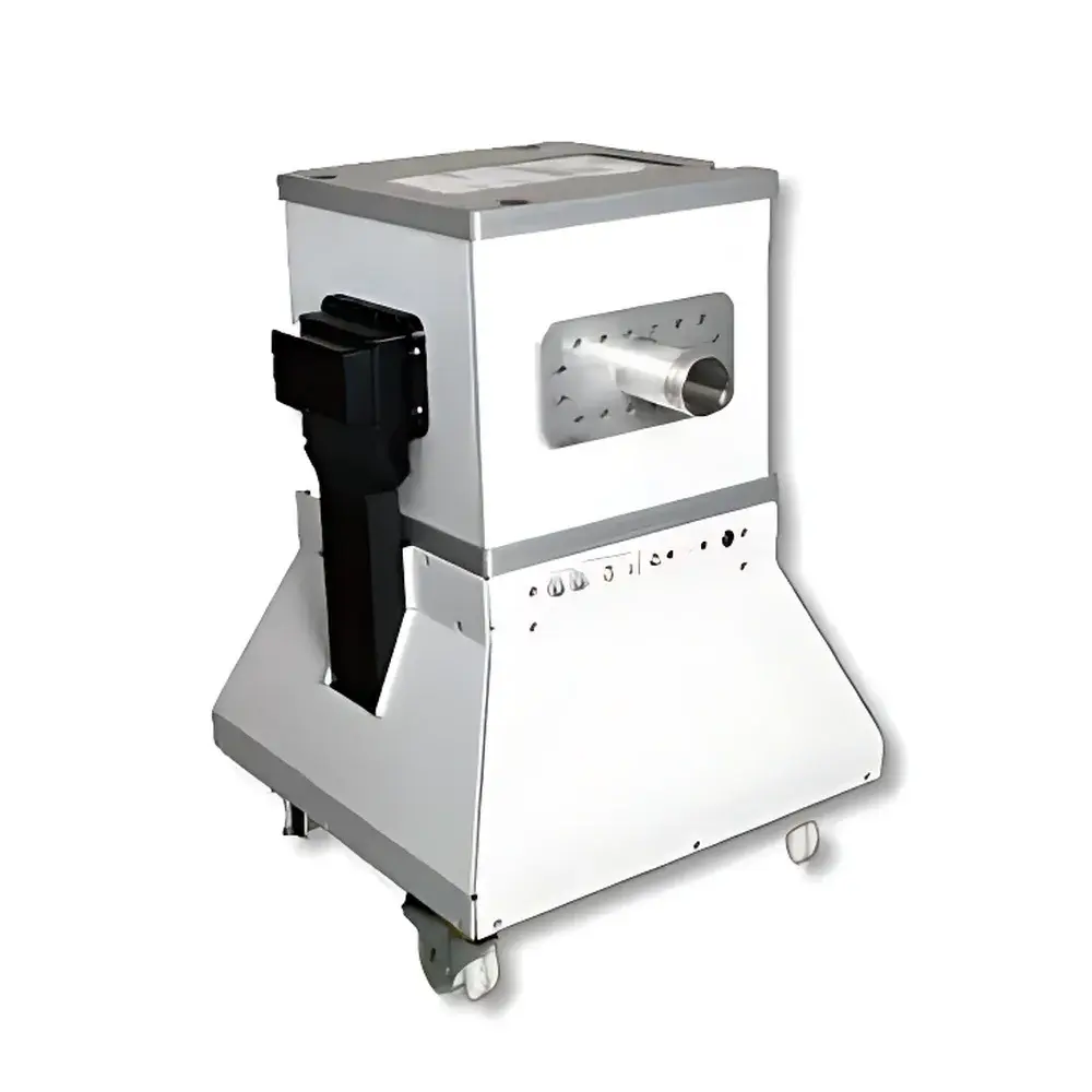
NIUMAG Aspect M3 Small Animal MRI System
| Brand | NIUMAG |
|---|---|
| Origin | Israel |
| Manufacturer Type | Authorized Distributor |
| Origin Category | Imported |
| Model | M3 |
| Instrument Type | Nuclear Magnetic Resonance (NMR) Imaging System |
| Magnet Type | Permanent Magnet, 1.0 Tesla |
| Field Homogeneity | <10 ppm over 20 mm DSV |
| Gradient Strength | ≥200 mT/m |
| Gradient Slew Rate | ≥1000 T/m/s |
| Spatial Resolution | ≤200 µm isotropic (typical in vivo mouse brain) |
| Scan Time | <10 minutes per standard sequence |
| Cooling | Passive (no cryogens or chiller required) |
| Shielding | Self-shielded, zero fringe field beyond instrument footprint |
| Regulatory Compliance | CE Marked, IEC 61000-6-3/6-4, ISO 13485–certified manufacturing processes |
| Software Platform | Aspect ImageSuite v5.x (FDA 21 CFR Part 11 compliant audit trail optional) |
Overview
The NIUMAG Aspect M3 Small Animal MRI System is a compact, self-shielded, permanent-magnet-based nuclear magnetic resonance imaging platform engineered exclusively for high-fidelity in vivo murine phenotyping. Operating at a stable 1.0 Tesla field strength, the system leverages optimized gradient performance (≥200 mT/m) and advanced RF coil architecture to deliver robust signal-to-noise ratio (SNR) and spatial fidelity in longitudinal studies. Unlike superconducting systems requiring liquid helium and active shielding, the M3 employs a passively cooled, maintenance-free permanent magnet assembly—eliminating cryogen dependency, reducing total cost of ownership, and enabling installation within standard laboratory spaces, including biosafety level 2 (BSL-2) containment rooms without structural reinforcement. Its homogeneous field (<10 ppm over 20 mm DSV) supports reproducible quantitative imaging across anatomical, functional, and contrast-enhanced protocols, making it suitable for preclinical research governed by GLP-compliant workflows.
Key Features
- Compact permanent magnet design (1.0 T), occupying <1.2 m² floor space with integrated RF shield
- Zero fringe field configuration: no external magnetic shielding or facility modifications required
- Fully passive thermal management: no cryogens, chillers, or scheduled magnet maintenance
- Dedicated mouse-specific RF coils (volume and surface arrays) with automatic tuning and matching
- Preconfigured pulse sequences optimized for murine anatomy: spin echo (SE), fast spin echo (FSE), gradient echo (GRE), and inversion recovery (IR)
- Integrated physiological monitoring interface (respiratory gating, ECG synchronization, temperature feedback)
- Modular hardware architecture supporting future upgrades including diffusion-weighted imaging (DWI) and MR spectroscopy (MRS) capability
Sample Compatibility & Compliance
The Aspect M3 accommodates live mice (typically 20–35 g) under isoflurane anesthesia via a dedicated stereotactic cradle with integrated heating and gas delivery. The system complies with ISO 13485–aligned quality management standards for medical device distribution and meets electromagnetic compatibility (EMC) requirements per IEC 61000-6-3 (emissions) and IEC 61000-6-4 (immunity). While not classified as a clinical diagnostic device, its acquisition protocols align with ASTM E2790 (Standard Guide for Preclinical MRI Protocol Development) and support data traceability frameworks required for regulatory submissions (e.g., FDA IND-enabling toxicology studies). Audit trail functionality—including user logins, parameter changes, and image export history—is available under optional 21 CFR Part 11 compliance mode.
Software & Data Management
Aspect ImageSuite v5.x provides a unified interface for sequence programming, real-time reconstruction, and quantitative analysis. The software includes DICOM 3.0 export, NIfTI conversion, and built-in tools for region-of-interest (ROI) quantification, T1/T2 mapping, and volumetric segmentation. All acquisitions are timestamped and associated with metadata (animal ID, anesthesia duration, coil type, sequence parameters), ensuring FAIR (Findable, Accessible, Interoperable, Reusable) data principles. Raw k-space data is preserved in vendor-neutral format for third-party processing (e.g., FSL, SPM, AFNI). ImageSuite supports automated batch processing pipelines and integrates with institutional PACS or LIMS via configurable DICOM SCU/SCP endpoints.
Applications
- Longitudinal tumor growth and metastasis monitoring (subcutaneous, orthotopic, and metastatic models)
- Neuroanatomical phenotyping: hippocampal volumetry, cortical thickness mapping, white matter integrity assessment
- Cerebrovascular imaging: cerebral blood volume (CBV), perfusion-weighted imaging (PWI), and stroke model validation
- Cardiac function analysis: cine-MRI for ejection fraction, wall motion, and chamber dimensions in transgenic models
- Metabolic disease phenotyping: hepatic steatosis quantification, pancreatic islet morphology, adipose tissue distribution
- Stem cell tracking using iron oxide–based contrast agents (e.g., USPIO)
- Bone microarchitecture assessment via ultra-short TE (UTE) sequences in osteoporosis and fracture-healing models
FAQ
Does the M3 require liquid helium or external cooling infrastructure?
No. The permanent magnet operates at ambient temperature with passive thermal stabilization—zero cryogens, chillers, or routine magnet servicing.
Can the system be installed inside a biosafety cabinet or BSL-2 room?
Yes. Its self-shielded design produces no measurable fringe field beyond the instrument enclosure, permitting placement behind biological containment barriers without compromising safety or image quality.
Is quantitative T1/T2 mapping supported out of the box?
Yes. Predefined inversion recovery and multi-echo spin echo protocols generate pixel-wise T1 and T2 maps with automated fitting algorithms and uncertainty estimation.
What level of regulatory compliance does the software support?
ImageSuite v5.x offers optional 21 CFR Part 11–compliant audit trail, electronic signatures, and role-based access control—validated for use in GLP and GCP environments.
How is image resolution validated for murine applications?
Resolution is verified using NIST-traceable phantom inserts (e.g., ACR MRI Phantom Model 604); typical in vivo isotropic resolution is 195–235 µm depending on sequence and SNR constraints.


