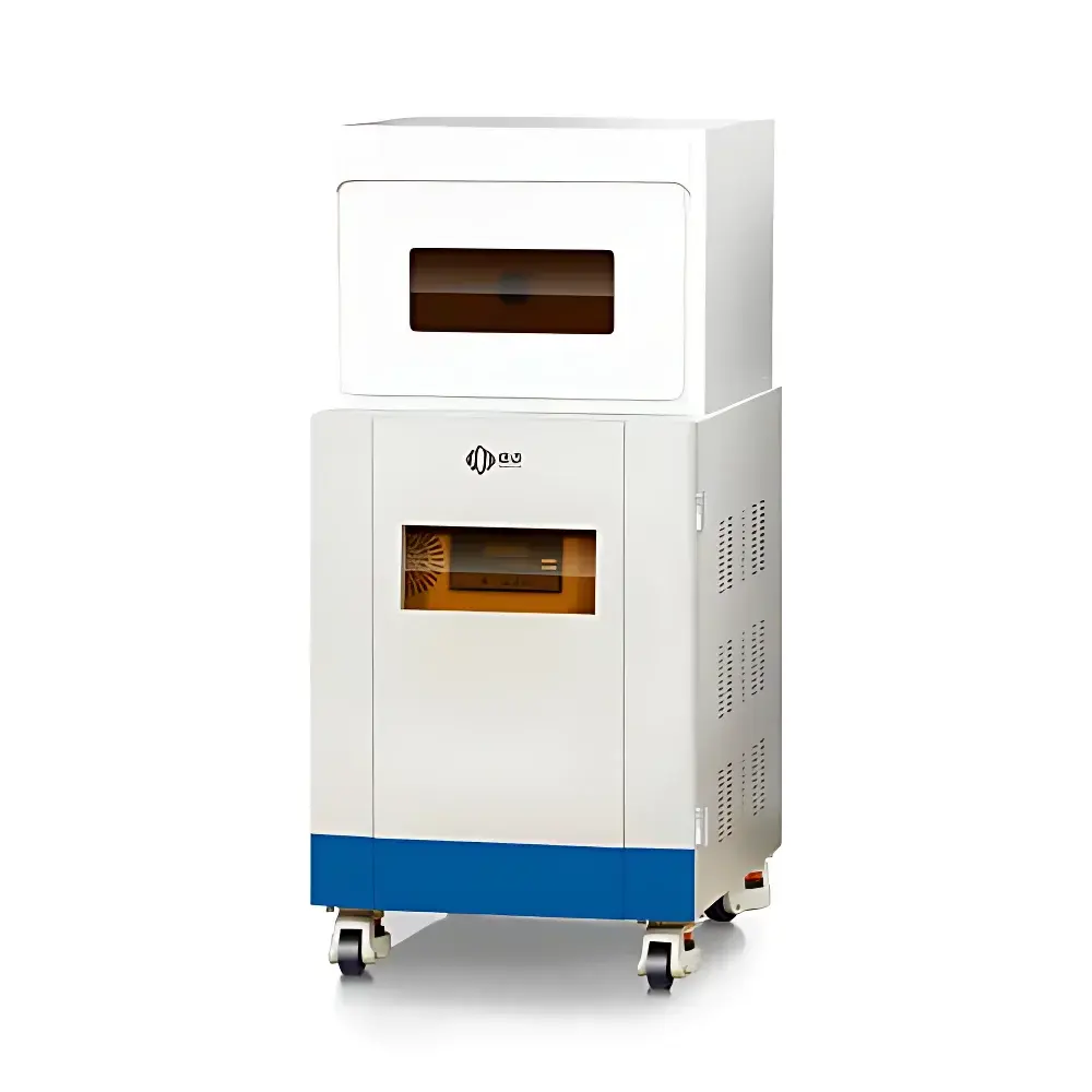
NIUMAG NM21-040H-I Small Animal MRI System
| Brand | NIUMAG |
|---|---|
| Origin | Shanghai, China |
| Manufacturer Type | Authorized Distributor |
| Region of Manufacture | Domestic (China) |
| Model | NM21-040H-I |
| Instrument Type | Benchtop Permanent-Magnet MRI System |
| Magnetic Field Strength | 0.5 ± 0.08 T |
| RF Coil Inner Diameter | 40 mm |
| Field of View (FOV) | 60 × 60 mm² |
| Maximum Sample Capacity | 1 animal per scan |
| Energy Resolution | Not applicable (N/A) |
| Spatial Resolution | Not specified in source |
| Scan Speed | Not quantified (system supports multi-sequence acquisition with variable TR/TE) |
| Compliance | Designed for preclinical GLP-compliant research environments |
Overview
The NIUMAG NM21-040H-I is a compact, permanent-magnet-based benchtop magnetic resonance imaging (MRI) system engineered specifically for non-invasive, high-contrast structural and functional imaging of small laboratory animals. Operating at a static magnetic field strength of 0.5 ± 0.08 T, the system leverages robust permanent magnet architecture to deliver stable field homogeneity without cryogens or active shimming infrastructure—significantly reducing operational overhead and facility requirements. Its 40 mm inner-diameter radiofrequency (RF) gradient coil accommodates standard murine models including C57BL/6 mice, BALB/c nude mice, and juvenile rats, enabling reproducible longitudinal studies within constrained vivarium or core imaging lab spaces. Unlike superconducting MRI platforms, the NM21-040H-I requires no liquid helium refills, no water-cooling circuits, and minimal site preparation—making it suitable for installation in standard laboratory rooms with standard power supply (220 V AC, 50 Hz). The system employs spin-echo and gradient-echo pulse sequences to generate T1-, T2-, and proton density-weighted contrast, supporting anatomical delineation of soft-tissue structures such as brain parenchyma, liver lobules, renal cortex, and subcutaneous tumor xenografts.
Key Features
- Permanent magnet design ensures zero cryogen consumption and eliminates quench risk—ideal for shared core facilities and teaching laboratories.
- Integrated gradient subsystem with X/Y/Z linearity >90% over 40 mm DSV (Diameter Spherical Volume), optimized for high-fidelity spatial encoding in small-animal applications.
- Dedicated 40 mm RF volume coil provides uniform B1 excitation and signal reception across typical mouse head and torso dimensions.
- Field of view configurable up to 60 × 60 mm² with isotropic voxel resolution adaptable via matrix size and bandwidth selection—enabling trade-offs between SNR, resolution, and acquisition time.
- Thermally stabilized magnet housing maintains field drift < 50 ppm/hour over ambient temperature fluctuations (18–25 °C), ensuring measurement repeatability across multi-day experiments.
- Modular software interface supports DICOM export, ROI-based quantitative analysis, and sequence parameter scripting for custom protocol development.
Sample Compatibility & Compliance
The NM21-040H-I is validated for use with live, anesthetized rodents under IACUC-approved protocols. Compatible sample types include: adult and neonatal mice (up to 35 g), nude mice, and juvenile Sprague-Dawley rats (≤100 g). Anesthesia integration (isoflurane-compatible gas delivery port) and physiological monitoring inputs (respiratory gating, temperature feedback) are supported via optional accessories. The system complies with electromagnetic emission limits per CISPR 11 Group 1 Class B and meets mechanical safety standards per ISO 13857. While not FDA-cleared for human use, it adheres to principles outlined in ISO/IEC 17025 for test equipment validation and supports audit-ready documentation for GLP-regulated preclinical studies—including timestamped raw data archiving and user-access logs.
Software & Data Management
Acquisition and reconstruction are managed through NIUMAG’s proprietary MRStudio platform, compatible with Windows 10 (64-bit) and featuring a graphical sequence editor, real-time k-space visualization, and automated shimming routines. All image datasets are stored in vendor-neutral NIfTI format and exported to DICOM 3.0 Level 3 (Enhanced MR Image Storage) for integration into PACS or third-party analysis pipelines (e.g., FSL, SPM, ITK-SNAP). Audit trail functionality records operator ID, acquisition parameters, timestamps, and software version—supporting compliance with 21 CFR Part 11 when deployed with validated electronic signature modules. Raw k-space data can be exported for offline reconstruction using MATLAB or Python-based toolkits (e.g., SigPy, BART).
Applications
- Longitudinal monitoring of orthotopic and subcutaneous tumor growth kinetics and treatment response.
- Neuroimaging of stroke models (MCAO), neurodegeneration (APP/PS1), and traumatic brain injury.
- Hepatic fat quantification (PDFF mapping) and fibrosis staging in NAFLD/NASH models.
- Renal morphometry and perfusion assessment in diabetic nephropathy studies.
- In vivo evaluation of nanoparticle-based MRI contrast agents—including relaxivity (r1/r2) characterization and biodistribution kinetics.
- Cardiac cine MRI for left ventricular ejection fraction (LVEF) and wall motion analysis in murine heart failure models.
- Embryonic development tracking via high-resolution static imaging of ex utero cultured embryos (E12.5–E18.5).
FAQ
Is the NM21-040H-I capable of functional MRI (fMRI) or diffusion-weighted imaging (DWI)?
Yes—DWI is supported via pulsed-gradient spin-echo sequences; fMRI (BOLD contrast) is feasible with respiratory-gated EPI, though SNR limitations apply at 0.5 T.
What anesthesia systems are compatible?
The system includes standardized ports for integration with commercial isoflurane vaporizers (e.g., SomnoSuite, Euthanex) and supports nose-cone or whole-body chamber delivery.
Does the system support multi-slice or 3D volumetric acquisition?
Yes—both 2D multi-slice and 3D gradient-echo acquisitions are implemented, with slice thickness adjustable from 0.5 mm to 2.0 mm.
Can data be processed externally using open-source tools?
Absolutely—raw k-space data and reconstructed NIfTI volumes are fully accessible and documented for interoperability with AFNI, FSL, and Python-based neuroimaging libraries.
What maintenance schedule is recommended?
Annual field homogeneity verification and RF coil performance calibration are advised; no magnet servicing is required due to permanent magnet construction.


