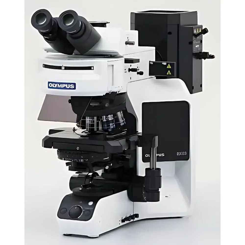
Olympus BX53 Fluorescence Microscope
| Brand | Olympus |
|---|---|
| Origin | Japan |
| Instrument Type | Upright Biological Microscope |
| Model | BX53 |
| Illumination | 100 W Mercury Arc Lamp (Fluorescence), Halogen/LED (Transmitted Light) |
| Objective Turret | Manual or Motorized 7-Position |
| Stage | Mechanical Right-Hand Control, 76 × 52 mm Travel |
| Condenser | Motorized Universal Condenser (NA 0.9–1.4, WD 0.63–2.0 mm) |
| Observation Modes | Brightfield, Darkfield, Phase Contrast, Simple Polarization, DIC, Fluorescence (UV, Blue, Green Excitation) |
| Eyepiece Field Number | FN 22 or FN 26.5 |
| Dimensions (W×D×H) | 274.5 × 614 × 469 mm |
| Weight | 21 kg (Fluorescence Configuration) |
Overview
The Olympus BX53 Fluorescence Microscope is an upright, research-grade optical platform engineered for high-fidelity multicontrast imaging in life science laboratories. Built upon Olympus’ proven UIS2 optical system, the BX53 integrates precision mechanical architecture with advanced fluorescence optics to support quantitative imaging across brightfield, phase contrast, differential interference contrast (DIC), darkfield, simple polarization, and multi-band fluorescence modalities. Its optical design adheres to Köhler illumination principles for both transmitted and epi-illumination paths, ensuring uniform intensity distribution and minimal photometric drift—critical for reproducible fluorescence quantification and time-lapse in vivo imaging. The microscope’s modular chassis accommodates motorized and encoded components without compromising mechanical stability, making it suitable for GLP-compliant histopathology workflows, cell biology assays, and developmental biology studies requiring long-term specimen observation under controlled environmental conditions.
Key Features
- UIS2 X-Line objective series: Delivers flat-field correction across FN 22–26.5, enhanced chromatic aberration correction (including violet and near-UV wavelengths), and high NA (up to 1.45) for superior resolution and signal-to-noise ratio in weak-fluorescence applications.
- Dedicated fluorescence illumination system: Equipped with a 100 W mercury arc lamp and optional 75 W xenon source; features an 8-position excitation filter turret with high-transmission, steep-edge dichroic mirrors (>99% stray light suppression) and integrated fly-eye lens for uniform epi-illumination across the full field of view.
- Motorized universal condenser: Supports NA ranging from 0.9 to 1.4 with adjustable top lens swing-out and automatic iris closure—minimizing back-reflection and autofluorescence during fluorescence acquisition.
- Ergonomic stage and focusing mechanism: Ceramic-coated mechanical stage with 76 × 52 mm travel and low-friction steel-cable drive; optional right-hand control lever with rubberized grip and height-adjustable handle extension reduces operator fatigue during extended sessions.
- Encoded hardware options: Motorized objective turret, excitation filter wheel, and condenser enable automatic metadata logging (magnification, contrast mode, exposure parameters) directly into acquisition software—supporting 21 CFR Part 11–compliant audit trails when paired with Olympus cellSens software.
Sample Compatibility & Compliance
The BX53 accommodates standard 1–3 mm thick glass slides, coverslips (0.13–0.17 mm), and live-cell chambers (e.g., µ-Slide 8-well, Lab-Tek chambered coverglasses). Its wide working distance condensers (WD ≥ 0.63 mm) and oil-immersion-capable objectives support high-resolution imaging of tissue sections, fixed and live mammalian cells, zebrafish embryos, and C. elegans specimens. The system complies with ISO 10934-1 (microscope nomenclature), ISO 8578 (mechanical stability), and IEC 61000-6-3 (EMC emissions). When configured with encoded components and validated cellSens software, it meets documentation requirements for FDA-regulated preclinical imaging workflows and supports traceable calibration per ASTM E2858 (quantitative fluorescence microscopy).
Software & Data Management
Olympus cellSens software provides native integration with BX53 hardware for automated acquisition, multi-channel registration, Z-stack reconstruction, and intensity-based colocalization analysis. The software supports TIFF, ND2, and OME-TIFF export formats compliant with Bio-Formats and OMERO repositories. Audit trail functionality logs user actions, parameter changes, and hardware state transitions—including objective selection, excitation filter position, and condenser NA setting—with timestamped, non-erasable records. Raw image data retains embedded EXIF metadata (exposure time, gain, lamp intensity, magnification), enabling retrospective validation and cross-platform reanalysis. Optional cellSens Entry and cellSens Dimension licenses accommodate tiered workflow needs—from routine pathology documentation to publication-ready 3D rendering.
Applications
- Diagnostic histopathology: Simultaneous brightfield and fluorescence assessment of immunohistochemically stained tissue sections using dual-modality observation tubes.
- Live-cell dynamics: Long-term DIC + fluorescence time-lapse of subcellular structures (e.g., mitochondria, lysosomes) under temperature- and CO₂-controlled stages.
- FISH and multicolor genomics: High-sensitivity detection of ≤1 kb DNA probes via optimized UV/blue/green excitation channels and narrow-band emission filters.
- Neuroscience: High-NA imaging of dendritic spines and synaptic puncta in brain slice preparations, leveraging DIC contrast for morphology and fluorescence for molecular labeling.
- Microbial analysis: Phase contrast + fluorescence screening of bacterial biofilms or GFP-expressing strains on agarose pads or microfluidic devices.
FAQ
Does the BX53 support automated focus mapping for Z-stack acquisition?
Yes—when equipped with a motorized fine-focus drive and compatible cellSens software, the BX53 enables programmable Z-series acquisition with sub-micron step resolution and autofocus compensation between slices.
Can the BX53 be integrated into a GMP-compliant imaging workflow?
Yes—through use of encoded hardware, electronic signatures, and audit-trail-enabled cellSens software, the system satisfies core elements of FDA 21 CFR Part 11 and EU Annex 11 for regulated environments.
What is the maximum usable magnification with oil immersion objectives?
The BX53 supports UIS2 100× oil immersion objectives (NA 1.45), delivering theoretical resolution down to 200 nm at 550 nm wavelength, consistent with Abbe diffraction limits.
Is LED illumination available for fluorescence excitation?
While the standard fluorescence illumination uses mercury or xenon arcs, Olympus offers LED-based excitation modules (e.g., X-Cite Exacte) as third-party-compatible upgrades—providing stable, cool operation and extended lifetime without spectral compromise.
How does the BX53 minimize autofluorescence in transmitted-light fluorescence imaging?
By combining high-transmission, low-autofluorescence UIS2 objectives with motorized condenser positioning (top lens retracted, iris closed), the system suppresses background signal from glass substrates and mounting media—critical for detecting dim endogenous fluorophores such as NAD(P)H or collagen SHG signals.
Related Products


