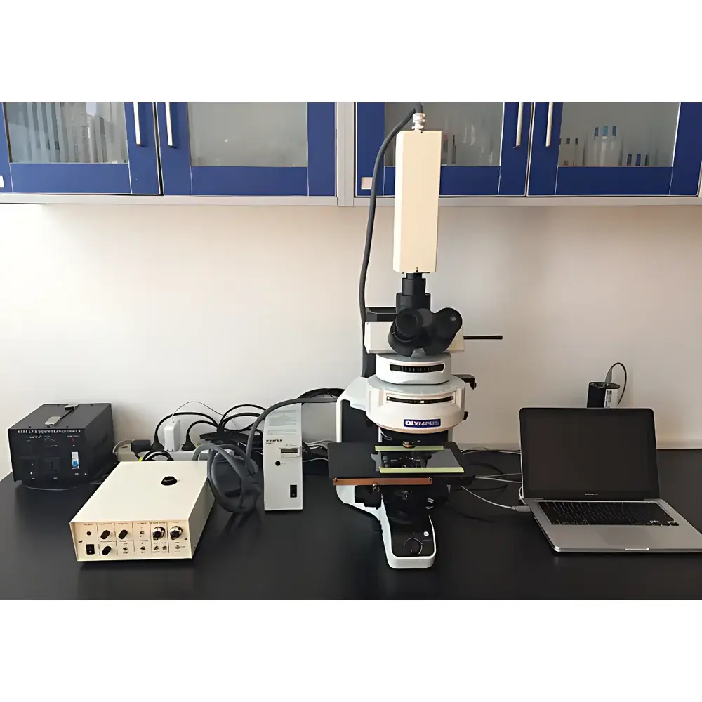
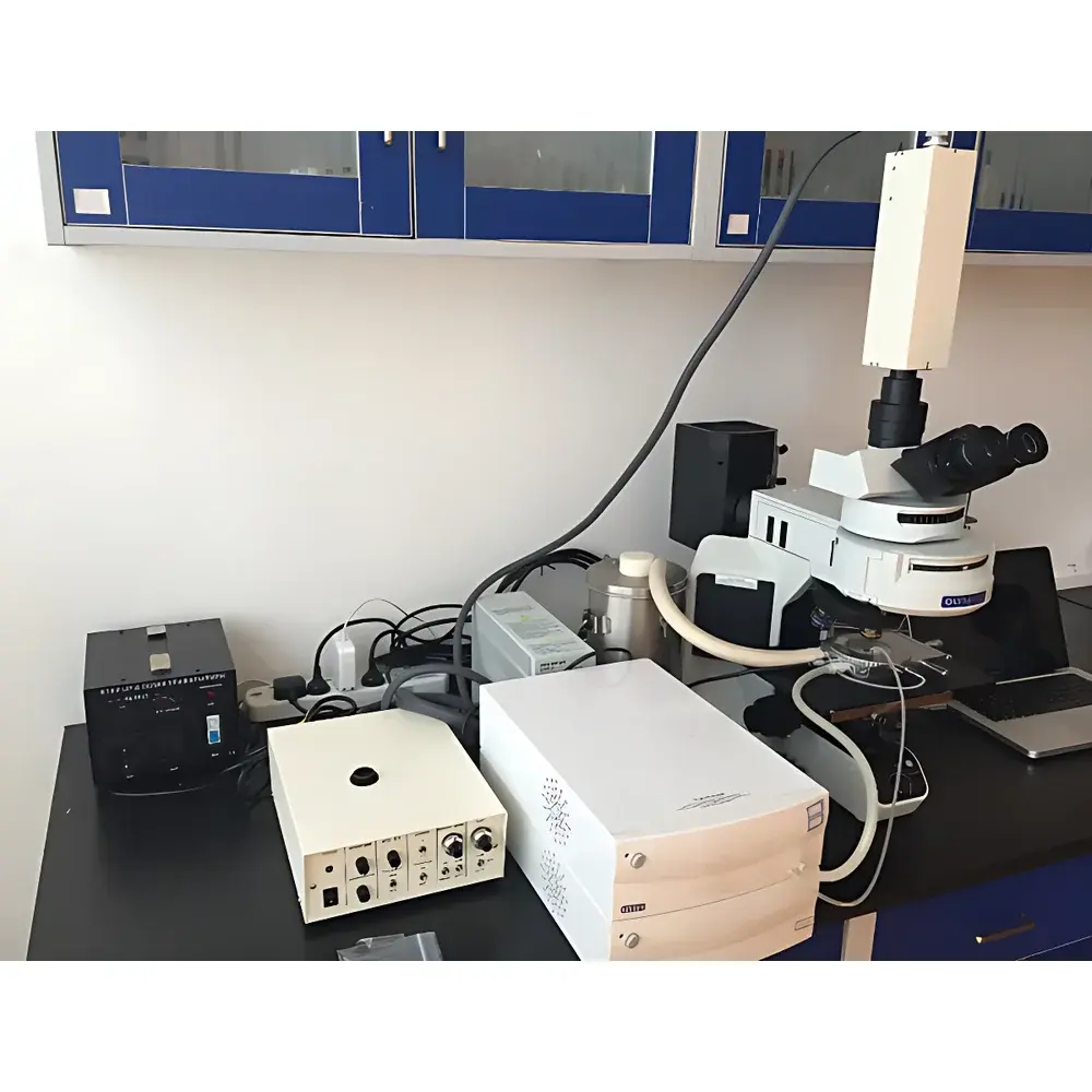
Olympus BX53-IR Infrared Microscope System
| Origin | USA |
|---|---|
| Manufacturer Type | Authorized Distributor |
| Origin Category | Imported |
| Model | BX53-IR |
| Pricing | Upon Request |
Overview
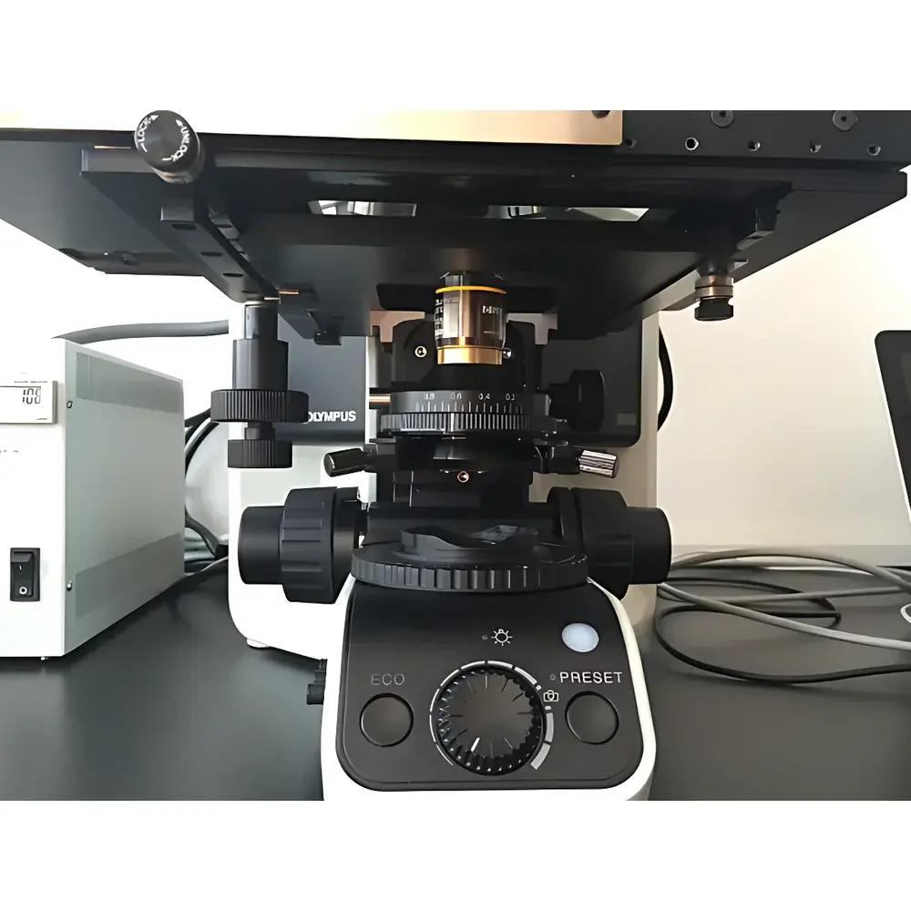
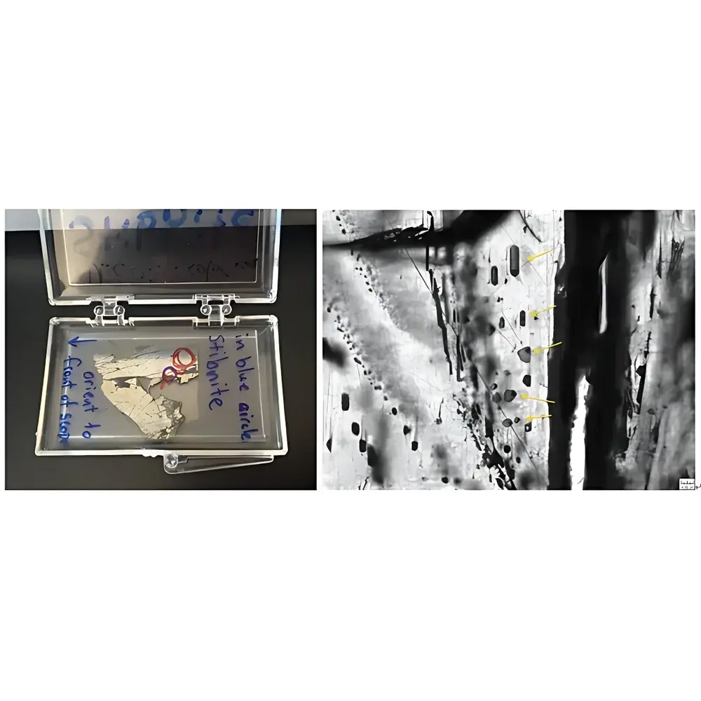
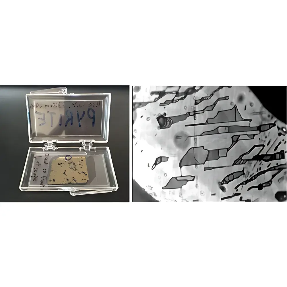
The Olympus BX53-IR Infrared Microscope System is a purpose-engineered analytical platform designed for high-fidelity fluid inclusion characterization in opaque and semi-transparent minerals. Unlike conventional optical microscopes, this system integrates three optically isolated illumination pathways—visible-light transmission, epifluorescence reflection, and modified mid-infrared (IR) transmission—enabling correlative multimodal analysis across 400–2200 nm. Its core measurement principle relies on IR transmittance contrast imaging: infrared radiation penetrates optically opaque matrices (e.g., pyrite, stibnite) where visible light is fully absorbed, revealing internal fluid inclusions via differential absorption and scattering. The system’s IR transmission path is mechanically and optically optimized using a custom-modified condenser assembly, wherein a 10× objective serves as the condenser lens—enhancing numerical aperture, increasing irradiance at the sample plane, and improving signal-to-noise ratio for weakly transmitting mineral hosts.
Key Features
- Triple-path optical architecture: simultaneous visible-light transmission (400–700 nm), UV/visible epifluorescence (365–488 nm excitation), and broadband IR transmission (up to 2200 nm)
- Custom-modified BX53-IR microscope frame with IR-optimized optics, including CaF2-lens objectives and anti-reflection coated IR-transmissive beam splitters
- Dual-camera imaging subsystem: Q-Imaging R2000 high-speed digital camera for visible/fluorescence/NIR (≤1100 nm); analog IR camera with InSb or MCT sensor for extended NIR-SWIR (1100–2200 nm)
- Integrated THMSG600 heating-cooling stage with T95 precision temperature controller and LN95 liquid nitrogen cooling unit, enabling in-situ thermal analysis from –196 °C to +600 °C with ±0.2 °C stability
- Motorized XYZ stage with 100 nm repeatability and software-controlled focus mapping for automated inclusion localization and thermal profiling
Sample Compatibility & Compliance
The system accommodates standard 25 mm and 32 mm polished thin sections, doubly polished wafers, and epoxy-mounted grain mounts. It supports both reflective and transmissive IR observation modes, making it suitable for sulfides (e.g., pyrite, chalcopyrite), oxides (e.g., hematite, magnetite), and semi-transparent sulfosalts (e.g., stibnite, realgar). All optical components comply with ISO 10110-7 (optical element surface quality) and ASTM E2339 (standard practice for microscopical examination of minerals). Temperature control hardware meets IEC 61000-4-3 (EMC immunity) and UL 61010-1 safety requirements. Data acquisition workflows support audit trails compliant with FDA 21 CFR Part 11 when paired with validated LIMS integration.
Software & Data Management
Control and analysis are performed via Olympus cellSens Dimension software (v3.1+), configured with custom IR acquisition modules. The software enables synchronized multi-channel image capture, spectral registration, temperature-stamped time-lapse recording, and geometric calibration for quantitative inclusion size and spatial distribution analysis. Raw data are stored in vendor-neutral TIFF and HDF5 formats, preserving metadata (wavelength, temperature, objective ID, stage coordinates). Export options include CSV for statistical post-processing and OME-TIFF for interoperability with open-source platforms such as ImageJ/Fiji and Python-based scikit-image pipelines. Audit logs record user actions, parameter changes, and instrument state transitions—fully traceable for GLP/GMP documentation.
Applications
- In-situ microthermometry of fluid inclusions in opaque ore minerals for hydrothermal deposit genesis modeling
- Phase identification and homogenization temperature (Th) determination in CO2-H2O-NaCl systems within pyrite-hosted inclusions
- Discrimination of hydrocarbon-bearing vs. aqueous inclusions via fluorescence-IR correlation mapping
- Thermal stability assessment of metastable phases (e.g., clathrates, hydrates) under controlled cryo-heating cycles
- Quantitative textural analysis of inclusion assemblages for geobarometry and fluid evolution reconstruction
FAQ
What wavelength range does the IR imaging subsystem cover?
The system supports dual-band IR imaging: 400–1100 nm using the Q-Imaging R2000 sCMOS camera, and 1100–2200 nm using a cooled InSb analog IR camera.
Can the THMSG600 stage be used for freezing-point depression measurements?
Yes—the LN95 cooling module provides stable sub-zero operation down to –196 °C, enabling eutectic temperature determination and ice-melting point analysis in saline inclusions.
Is the BX53-IR compatible with third-party spectroscopy add-ons?
The trinocular port and standardized C-mount interfaces allow integration with FTIR spectrometers or dispersive NIR spectrometers for point-spectral acquisition on individual inclusions.
Does the system support automated inclusion mapping across large-area sections?
Yes—motorized stage control, autofocus routines, and scripting API in cellSens enable unattended grid-based scanning and coordinate-tagged inclusion database generation.
Are optical components certified for use in cleanroom environments?
All IR-transmissive optics are cleaned to Class 100 (ISO 5) standards and supplied with contamination-controlled packaging; optional quartz-housed stages available for ISO 14644-1 Class 5 compliance.


