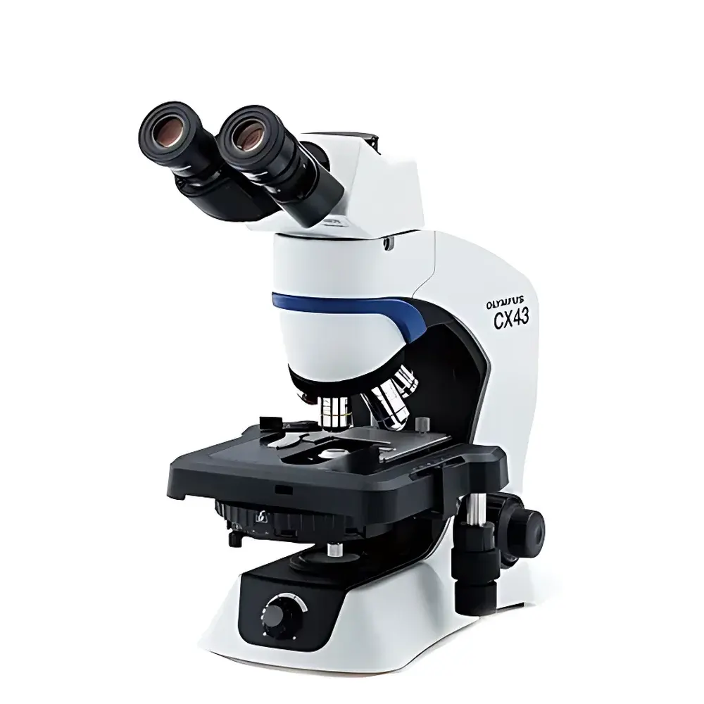
OLYMPUS CX43 Biological Microscope
| Brand | OLYMPUS |
|---|---|
| Origin | Japan |
| Model | CX43 |
| Microscope Type | Upright Biological Microscope |
| Optical System | UIS2 Infinity-Corrected Optics |
| Illumination | Integrated LED Köhler Illumination (2.4 W, Daylight Color Temperature ~5,500 K) |
| Objective Turret | Fixed 5-Position Nosepiece |
| Stage | Mechanical Stage (211 mm × 154 mm), X–Y Travel: 76 mm × 52 mm |
| Eyepieces | Widefield 10× (Field Number 20 mm) |
| Objectives | Plan Achromat UIS2 Series (2×, 4×, 10×, 20×, 40×, 60×, 100× Oil) |
| Contrast Methods Supported | Brightfield, Phase Contrast, Darkfield, Polarization, Fluorescence |
| Imaging Port | Trinocular C-Mount Output (1× or 0.5× optional) |
| LED Lifetime | ≥60,000 hours |
| Ergonomic Features | Low-Positioned Focus Knobs, Inclined Binocular/Trinocular Tube Options, Adjustable Stage Height & Eyepoint |
Overview
The OLYMPUS CX43 Biological Microscope is an upright, infinity-corrected optical platform engineered for routine and advanced life science applications in academic laboratories, clinical pathology departments, and quality control environments. Built upon the UIS2 optical system, it delivers flat-field, high-fidelity imaging across wide fields of view—essential for consistent morphological assessment, cell counting, histopathology screening, and live specimen documentation. Its LED-based Köhler illumination ensures uniform brightness and stable color temperature (~5,500 K) at all intensity levels, eliminating manual white-balance recalibration during dynamic observation or digital capture. Designed with ISO 9001-certified manufacturing standards in Japan, the CX43 integrates mechanical robustness with ergonomic intelligence: low-mounted coarse/fine focus controls, intuitive stage positioning, and minimized hand movement during specimen handling—all contributing to reduced operator fatigue during extended daily use.
Key Features
- Ergonomic Human-Centered Design: Focus knobs positioned at optimal height allow users to rest forearms on the stage while adjusting focus; stage control knobs are co-located with focus controls for coordinated X–Y–Z manipulation.
- UIS2 Infinity-Corrected Optics: Plan achromat objectives deliver diffraction-limited resolution and field flatness up to FN20, minimizing edge distortion and chromatic aberration across magnifications from 2× to 100× oil immersion.
- Stable LED Illumination System: 2.4 W daylight-balanced LED provides flicker-free, heat-free illumination with >60,000-hour rated lifetime—ensuring long-term photometric consistency without output decay or spectral shift.
- Modular Contrast Flexibility: Standard brightfield configuration supports seamless upgrade paths to phase contrast (with optional PH condenser), fluorescence (via rear-mounted compact LED fluorescence unit), polarization (CX3-KPA accessory), and darkfield (specialized condenser).
- Five-Position Objective Turret: Accommodates standard objectives plus specialized optics—including 2× macro objectives for overview scanning and long-working-distance phase contrast lenses—enabling multi-modal workflow consolidation on a single frame.
- Digital Imaging Readiness: C-mount trinocular port (1× or 0.5× reduction available) ensures precise alignment with scientific CMOS/CCD cameras; integrated shutter and exposure synchronization support GLP-compliant image acquisition protocols.
Sample Compatibility & Compliance
The CX43 accommodates standard glass microscope slides (up to 3 mm thickness), Petri dishes (with optional dish holder), and tissue culture flasks (using inverted-stage adapters). Its mechanical stage supports both single-slide clamping and dual-slide configurations, with calibrated X–Y verniers and travel limit stops for repeatable positioning. All optical components—including objectives and eyepieces—are coated with anti-fungal and hydrophobic layers compliant with JIS Z 8101-2:2015 for microbiological safety. The system meets IEC 61000-6-3 (EMC emissions) and IEC 61000-6-2 (immunity) standards. While not FDA 510(k)-cleared as a medical device, its optical performance aligns with ASTM E2872–21 (Standard Practice for Calibration of Microscope Magnification) and ISO 8578:2017 (Microscopes — Requirements for Biological Microscopes).
Software & Data Management
The CX43 operates independently of proprietary software but interfaces natively with third-party imaging platforms—including Olympus cellSens™ (v2.4+), ImageJ/Fiji, and HALO®—via standard USB 3.0 or GigE Vision protocols. When paired with Olympus DP28 or DP74 camera systems, metadata embedding (objective ID, magnification, exposure time, illumination intensity) complies with DICOM Supplement 145 (Digital Microscopy) and supports audit-trail generation per FDA 21 CFR Part 11 requirements when deployed in validated laboratory information management systems (LIMS). Optional U-EPA2 eyepoint adjusters and U-APT LED arrow pointers enable annotation-ready workflows for teaching, telepathology, and regulatory submissions.
Applications
- Routine hematology and urinalysis (RBC/WBC morphology, crystal identification using polarization)
- Histopathology slide screening and grading (e.g., Gleason scoring, mitotic index quantification)
- Microbiology colony isolation and Gram stain evaluation
- Cell culture monitoring (adherent and suspension lines) with optional phase contrast
- Fluorescent immunostaining validation (FITC/TRITC/DAPB channels with narrow-band excitation filters)
- Academic instruction and collaborative microscopy (dual-head viewing via optional tandem head)
FAQ
Is the CX43 compatible with oil immersion objectives?
Yes—the CX43 supports 100× oil immersion plan achromat objectives with spring-loaded retractable front lenses and integrated coverslip correction (0.17 mm).
Can the LED illumination be intensity-modulated during live-cell imaging?
Yes—continuous analog dimming (0–100%) is provided via front-panel rotary control or external TTL-compatible input for synchronized shutter triggering.
Does the CX43 meet GLP/GMP documentation requirements?
When integrated with compliant cameras and LIMS, the system supports traceable image metadata, electronic signatures, and audit logs—fulfilling core elements of GLP (OECD 1998) and GMP Annex 11 (EU) for non-clinical laboratory operations.
What is the maximum working distance for the 2× objective?
The UIS2 2× plan achromat offers a 140 mm working distance—ideal for large-volume specimens, Petri dishes, or stereo-level orientation prior to high-magnification examination.
Is service and calibration support available outside Japan?
OLYMPUS authorized service centers in North America, EMEA, and APAC provide ISO/IEC 17025-accredited calibration (magnification verification, resolution testing per ISO 9336-2), preventive maintenance, and optical alignment certification.
Related Products


