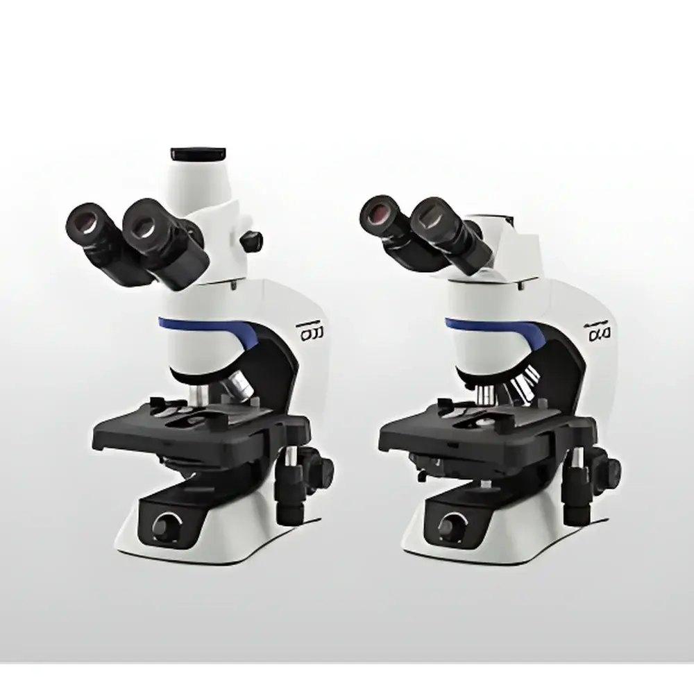
OLYMPUS CX43/CX33 Biological Microscope
| Brand | OLYMPUS |
|---|---|
| Origin | Japan |
| Manufacturer Type | Original Equipment Manufacturer (OEM) |
| Product Category | Imported Instrument |
| Model | CX43 / CX33 |
| Instrument Type | Upright Microscope |
| Illumination | Integrated LED with Köhler Illumination |
| Lifetime | 60,000 hours |
| Color Temperature Stability | Constant ~5,500 K across intensity range |
| Ergonomic Design | Low-positioned focus knobs, stage, and eyepoint optimization |
| Digital Imaging Interface | C-mount camera port |
Overview
The OLYMPUS CX43 and CX33 are upright biological microscopes engineered for high-throughput routine observation in academic teaching laboratories, clinical diagnostics, and quality control environments. Built upon OLYMPUS’ legacy of optical precision and mechanical reliability, these instruments employ a fixed Köhler illumination system combined with a stable, color-temperature-controlled white LED light source—designed to replicate natural daylight conditions (~5,500 K) across the full brightness range. This eliminates the need for repeated white-balance recalibration when adjusting illumination intensity, ensuring consistent specimen color fidelity during prolonged visual assessment or digital capture. The optical path is optimized for brightfield microscopy, supporting standard 10×–100× objective magnifications with parfocal, spring-loaded, and anti-fungal coated objectives. Both models feature a rigid, vibration-damped metal frame and a simplified optical train that prioritizes image clarity, contrast, and long-term alignment stability over modular complexity—making them ideal for standardized workflows where reproducibility, ease of use, and operator ergonomics are critical.
Key Features
- Ergonomic upright design with low-positioned coaxial focus knobs—enabling forearm support on benchtops and minimizing shoulder and wrist strain during extended use.
- Fixed Köhler illumination system: pre-aligned condenser and field diaphragm eliminate manual centering and focusing of the illumination path, reducing setup time and inter-user variability.
- Integrated high-stability LED light source with 60,000-hour rated lifetime; maintains constant color temperature (±50 K) from 10% to 100% intensity output—critical for comparative morphology analysis and color-sensitive staining protocols (e.g., H&E, Gram, Giemsa).
- Low-height mechanical stage with smooth, backlash-free X–Y translation and integrated specimen holder clips—optimized for rapid slide positioning and repeatable navigation under high-magnification objectives.
- Dual-port optical head configuration: standard trinocular tube with C-mount (1×) camera port supports integration with USB3.0 or CMOS-based digital imaging systems compliant with TWAIN/DCAM standards.
- Fine-focus limit stop mechanism prevents objective–coverslip collision during high-magnification (≥40×) focusing—protecting both expensive objectives and delicate live or stained specimens.
Sample Compatibility & Compliance
The CX43 and CX33 accommodate standard 1″ × 3″ (25 × 75 mm) glass microscope slides with coverslips (0.13–0.17 mm thickness), as well as petri dishes (up to 35 mm diameter) placed directly on the stage using optional dish holders. The microscope’s fixed working distance and standardized nosepiece threading (RMS 20.32 mm) ensure compatibility with third-party phase contrast, darkfield, or polarizing accessories certified to ISO 8578 and JIS B 7151. All optical components meet ISO 10934-1 (microscope nomenclature) and ISO 8578:2020 (mechanical interface specifications). The LED illumination system complies with IEC 62471 (photobiological safety) Class 1 requirements for continuous viewing. These instruments are routinely deployed in GLP-compliant histology labs and ISO 15189-accredited clinical pathology units where documentation of illumination consistency, focus repeatability, and stage positioning accuracy is required per internal SOPs.
Software & Data Management
While the CX43/CX33 operate as standalone optical instruments, their C-mount interface enables seamless integration with industry-standard imaging platforms—including OLYMPUS cellSens Entry (v2.4+), Media Cybernetics Image-Pro Premier, and open-source alternatives such as Fiji/ImageJ via compatible drivers. When paired with FDA 21 CFR Part 11–compliant acquisition software, the system supports audit-trail-enabled image capture, timestamped metadata embedding (objective ID, magnification, exposure, illumination intensity), and DICOM-SR export for PACS-integrated pathology workflows. Firmware updates (delivered via USB memory stick) maintain backward compatibility with legacy camera modules and preserve calibration integrity across instrument lifecycles.
Applications
- Routine hematology and urinalysis: rapid differential counts and crystal identification under brightfield at 40× and 100× (oil immersion).
- Microbiology training: Gram staining evaluation, yeast/bacterial morphology assessment, and motility observation without phase contrast add-ons.
- Plant and animal histology: section screening, tissue architecture verification, and immunohistochemistry (IHC) chromogen localization at consistent illumination conditions.
- Quality assurance in biomanufacturing: raw material inspection (e.g., cell culture contaminants), filter integrity testing, and sterility assay documentation.
- Undergraduate life science education: standardized microscopy modules aligned with ASBMB and HHMI curriculum benchmarks for optical competency and quantitative observation skills.
FAQ
Is the CX43/CX33 compatible with fluorescence observation?
No—these models are dedicated brightfield instruments. Fluorescence capability requires separate excitation filters, dichroic mirrors, and high-sensitivity detectors, which are supported only on OLYMPUS BX series platforms.
Can the LED illumination intensity be calibrated traceably?
Yes—the built-in photometric sensor allows relative intensity logging; for NIST-traceable calibration, users may integrate an external lux meter (e.g., Sekonic L-308S) at the eyepoint plane per ISO/IEC 17025 procedures.
What maintenance is required beyond routine cleaning?
Annual verification of Köhler alignment and LED output stability is recommended. No lamp replacement or condenser realignment is needed due to the fixed-optics architecture.
Does the microscope support motorized stage or autofocus?
No—motorization is not available on the CX platform. For automated scanning or z-stack acquisition, OLYMPUS recommends pairing with the BX63 or CKX53 systems.
Are replacement parts and service manuals available globally?
Yes—OLYMPUS provides multilingual service documentation, genuine spare parts (e.g., LED modules, eyepieces, objectives), and certified field service through authorized distributors in >90 countries under ISO 9001-certified support frameworks.


