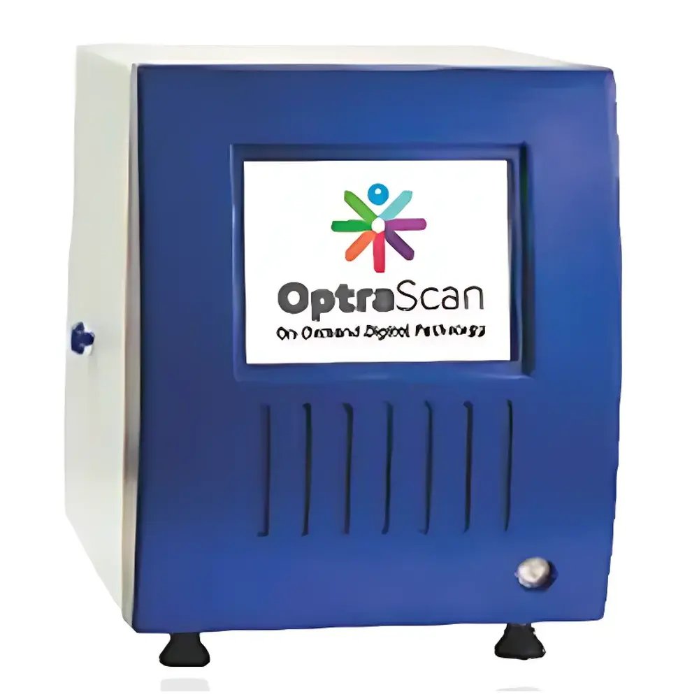
OptraScan Digital Slide Scanner System
| Brand | OptraScan |
|---|---|
| Origin | USA |
| Manufacturer Type | Authorized Distributor |
| Origin Category | Imported |
| Model | OptraScan |
| Pricing | Available Upon Request |
Overview
The OptraScan Digital Slide Scanner System is a compact, high-performance whole-slide imaging (WSI) platform engineered for precision digital pathology workflows in clinical laboratories, academic research institutions, and pharmaceutical development environments. Based on high-fidelity optical scanning architecture—combining motorized precision stage movement, multi-channel LED illumination, and sCMOS or CMOS sensor technology—the system captures full-resolution digital representations of glass histopathology slides with sub-micron spatial fidelity. It supports concurrent acquisition across brightfield (BF), fluorescence (FL), and confocal-capable modalities (depending on configuration), enabling standardized digitization of routine H&E-stained sections, immunohistochemistry (IHC)-processed specimens, fluorescence in situ hybridization (FISH) assays, and multiplexed biomarker panels. Designed for integration into ISO 15189- and CAP-accredited pathology labs, the OptraScan platform complies with DICOM-SR and HL7 FHIR standards for structured reporting and interoperability with enterprise PACS and LIS systems.
Key Features
- Modular hardware design accommodating OS-FL (fluorescence-optimized) and OS-FL/FLi (advanced fluorescence + AI-assisted analysis) configurations
- Automated slide loading with barcode recognition (1D/2D) and intelligent slide orientation detection
- Multi-spectral fluorescence excitation and emission filtering for simultaneous multi-probe FISH quantification (e.g., HER2/CEP17 ratio calculation per ASCO/CAP guidelines)
- High-throughput scanning: up to 40× objective resolution at ≤60 seconds per standard 15 mm × 15 mm tissue area in brightfield mode
- Optical path calibration with daily auto-focus and Z-stack acquisition support for thick-section or 3D tissue reconstruction
- Embedded GPU-accelerated preprocessing: flat-field correction, chromatic aberration compensation, and background subtraction
Sample Compatibility & Compliance
The OptraScan system accommodates standard 1″ × 3″ glass slides (up to 1 mm thickness), including charged, APES-coated, and electrostatically treated substrates. It is validated for use with formalin-fixed paraffin-embedded (FFPE) and frozen-section specimens stained via hematoxylin and eosin (H&E), immunoperoxidase (IHC), silver-enhanced in situ hybridization (SISH), and dual- or triple-color FISH protocols. Regulatory compliance includes adherence to FDA 21 CFR Part 11 requirements for electronic records and signatures when deployed with validated software modules; full audit trail logging, user role-based access control, and secure DICOM transmission are enabled by default. The system meets IEC 61010-1 safety standards and is CE-marked under the IVDR (In Vitro Diagnostic Regulation) framework for clinical diagnostic use in the EU.
Software & Data Management
OptraScan operates with proprietary OptraView™ software—FDA-cleared as a Class II medical device for primary diagnostic imaging in select jurisdictions. The software provides DICOM-compliant image storage, lossless compression (JPEG2000), and vendor-neutral export (SVS, NDPI, TIFF). Integrated analytical modules support quantitative pathology tasks: nuclear segmentation, membrane intensity scoring (e.g., HER2 IHC scoring per CAP guidelines), and FISH signal enumeration with automated centromere (CEP17) and locus-specific (HER2) probe counting. All processing logs—including scanner parameters, calibration timestamps, and user interventions—are retained in tamper-evident audit trails compliant with GLP and GCP documentation requirements. Cloud synchronization is implemented via HIPAA- and GDPR-compliant infrastructure, supporting encrypted remote review, collaborative annotation, and longitudinal case tracking across geographically distributed sites.
Applications
- Clinical diagnostics: Primary digital interpretation of prostate cancer biopsies (Gleason grading, tumor cell classification, stromal/tumor ratio quantification)
- Oncology biomarker validation: HER2 amplification status determination via FISH ratio analysis in breast and gastric carcinoma specimens
- Translational research: Multiplexed IHC/FISH co-localization studies for target engagement assessment in preclinical models
- Education and telepathology: Secure sharing of annotated WSI datasets for consensus review, proficiency testing, and remote second opinion
- Quality assurance: Digital archiving of reference slides for inter-laboratory comparison and CAP inspection readiness
FAQ
Does OptraScan support FDA-cleared diagnostic interpretation workflows?
Yes—OptraView™ software is FDA 510(k)-cleared (K211022) for primary diagnostic use in brightfield whole-slide imaging applications in the United States.
Can the system perform automated HER2/CEP17 ratio calculations required by CAP guidelines?
Yes—OS-FL and OS-FL/FLi models include validated FISH quantification algorithms that compute signal counts, classify nuclei, and report HER2/CEP17 ratios with configurable confidence thresholds and manual override capability.
Is DICOM-SR export supported for structured pathology reports?
Yes—structured reporting templates aligned with CAP Cancer Protocols and SNOMED CT terminology are embedded and exportable as DICOM Structured Reports.
What cybersecurity certifications apply to the cloud data service?
OptraScan Cloud Services are certified under ISO/IEC 27001:2022 and undergo annual third-party penetration testing; all data in transit and at rest is encrypted using AES-256 and TLS 1.3.
Are software updates and calibration services included in the maintenance agreement?
Yes—comprehensive service contracts include remote diagnostics, quarterly firmware/software releases, annual on-site optical calibration, and regulatory update notifications for evolving CLIA/CAP/ISO requirements.


