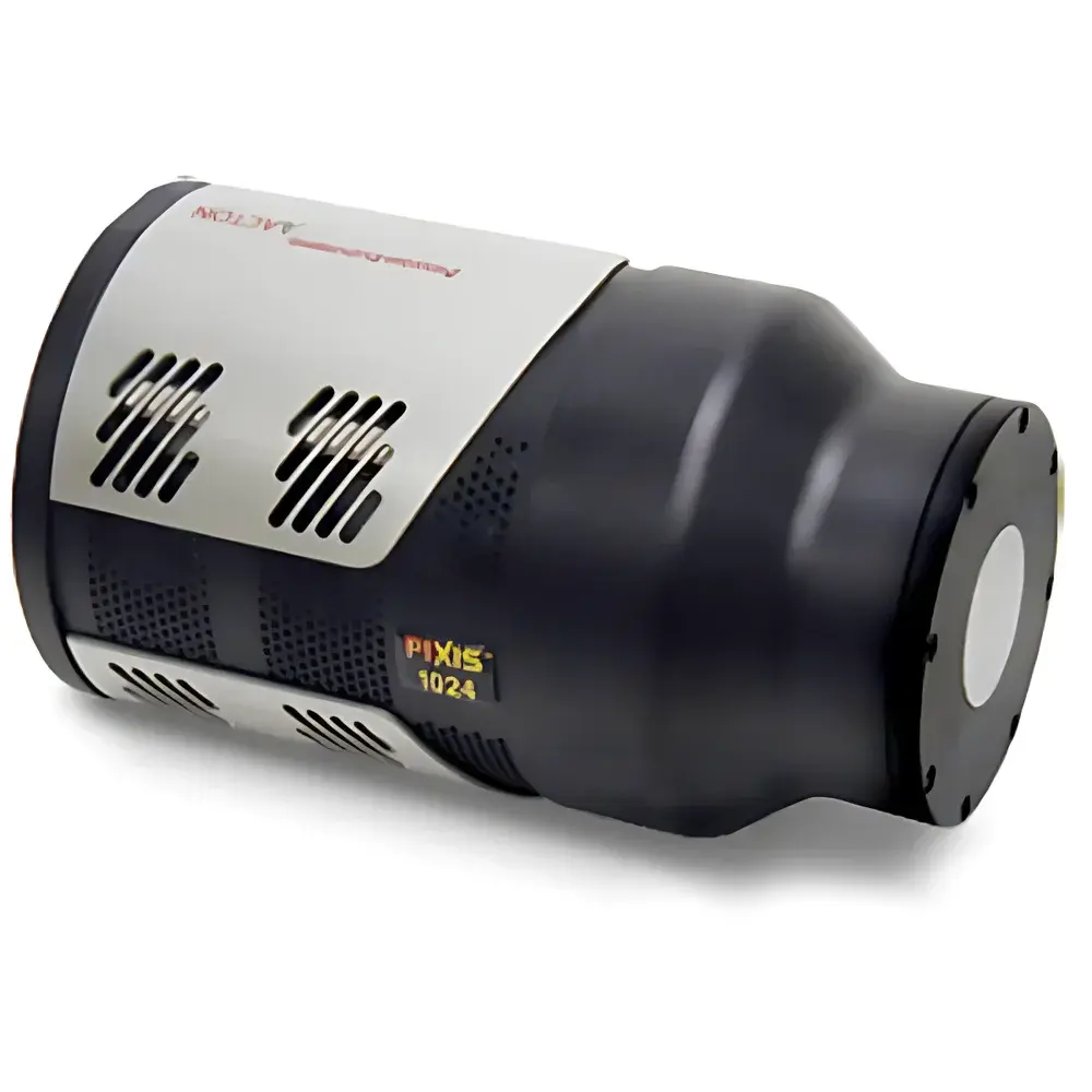
Princeton Instruments PIXIS-XF Indirect Detection X-ray Camera
| Brand | Teledyne Princeton Instruments |
|---|---|
| Origin | USA |
| Model Variants | XF-1024F, XF-1024B, XF-2048F, XF-2048B |
| Pixel Size | 13 × 13 µm (XF-1024 series), 13.5 × 13.5 µm (XF-2048 series) |
| X-ray Energy Range | <3 keV to >20 keV |
| Readout Speeds | 100 kHz and 2 MHz |
| ADC Resolution | 16-bit |
| Interface | High-speed USB 2.0 |
| Sensor Architecture | Front-illuminated (FI) or back-illuminated (BI) scientific CCD |
| Quantum Efficiency | Up to 97% at 550 nm (BI), 32% at 550 nm (FI) |
| Cooling | Thermoelectric (TE) with hot-spot suppression |
| Optical Coupling | Vacuum-integrated fiber-optic taper |
| Software Platform | LightField (64-bit), PICAM API |
Overview
The Princeton Instruments PIXIS-XF is a high-performance, indirect-detection X-ray imaging camera engineered for precision synchrotron, laboratory, and industrial applications requiring quantitative, low-noise, and energy-tunable detection across the soft to hard X-ray spectrum (sub-3 keV to >20 keV). Unlike direct-detection sensors, the PIXIS-XF employs a scintillator-based conversion architecture: incident X-rays are first absorbed by a replaceable phosphor screen—such as Gd₂O₂S:Tb—generating visible photons that are then efficiently coupled via a vacuum-integrated fiber-optic taper onto a scientific-grade CCD sensor. This design decouples radiation hardness requirements from the detector electronics while preserving spatial fidelity through 1:1 optical relay and enabling spectral optimization via screen selection. The system integrates ultra-low-noise, dual-gain readout electronics, thermoelectric cooling capable of stabilizing sensor temperature to ±0.1 °C, and a compact, maintenance-free mechanical housing suitable for integration into constrained beamlines or OEM platforms.
Key Features
- Modular scintillator interface: Rapid, in-situ replacement of phosphor screens (e.g., GdOS:Tb for 8–17 keV, CsI:Tl for lower energies) to optimize quantum detection efficiency across distinct X-ray energy bands
- Dual-readout amplifier architecture: Independent low-noise (100 kHz) and high-speed (2 MHz) channels support both high-dynamic-range imaging and real-time alignment/positioning tasks
- Scientific CCD options: Choice of front-illuminated (XF-1024F/XF-2048F) or back-illuminated (XF-1024B/XF-2048B) sensors, delivering peak QE up to 97% at 550 nm and full-well capacities exceeding 100,000 e⁻
- True 16-bit digitization: 100 kHz readout achieves <3 e⁻ rms read noise; 2 MHz mode maintains <15 e⁻ rms with full dynamic range preservation
- Vacuum-extended fiber-optic coupling: Minimizes optical loss and preserves MTF across the entire active area (100% fill factor); eliminates air-gap artifacts common in lens-coupled systems
- USB 2.0 plug-and-play interface: No frame grabber required; compatible with Windows 10/11 (64-bit) and Linux via PICAM SDK
Sample Compatibility & Compliance
The PIXIS-XF supports non-destructive, high-resolution radiography and tomography of diverse samples—from biological tissue sections and polymer composites to semiconductor wafers and geological cores. Its indirect detection scheme ensures compatibility with high-flux beamlines (e.g., APS, ESRF, SPring-8) and benchtop microfocus sources without sensor degradation. All models comply with IEC 61000-6-3 (EMC emission standards) and meet CE marking requirements for laboratory instrumentation. For regulated environments—including GLP-compliant preclinical imaging or FDA-submitted medical device characterization—the LightField software platform supports audit-trail logging, user-access controls, and 21 CFR Part 11–ready electronic signatures when deployed with validated configuration protocols.
Software & Data Management
LightField v8.x (64-bit) provides unified control of acquisition parameters, real-time image processing, and metadata embedding. Built-in routines include automatic dark-frame subtraction, flat-field correction using reference exposures, pixel defect mapping, and intensity linearity calibration traceable to NIST-certified photodiode standards. Raw data export supports HDF5, TIFF (16-bit), and FITS formats with embedded EXIF-like headers containing exposure time, sensor temperature, gain setting, and phosphor type. The PICAM Application Programming Interface enables seamless integration into Python, MATLAB, LabVIEW, or custom C/C++ acquisition frameworks—facilitating automated workflows in synchrotron shutter control loops or multi-modal correlative microscopy pipelines.
Applications
- Micro-CT & Phase-Contrast Imaging: Sub-micron resolution volumetric reconstruction enabled by high MTF (>40% at Nyquist) and sub-pixel registration stability over multi-hour scans
- X-ray Diffraction (XRD) & Coherent Diffractive Imaging (CDI): Low-noise, high-dynamic-range capture of weak Bragg peaks and speckle patterns; compatible with PILATUS-style hybrid pixel detectors for cross-validation
- Soft X-ray Microscopy (SXM): Optimized response below 1 keV using thin CsI:Tl screens; supports zone-plate illumination at wavelengths down to 2.4 nm (517 eV)
- Industrial NDT & Semiconductor Metrology: Real-time inspection of solder joint integrity, void detection in encapsulants, and metrology of EUV mask defects at 13.5 nm equivalent dose levels
- Time-Resolved Radiography: Synchronized triggering (TTL input/output) enables pump-probe studies with temporal resolution limited only by source pulse width and shutter latency
FAQ
What scintillator materials are supported, and how do they affect energy response?
Standard options include Gd₂O₂S:Tb (optimized for 8–17 keV), CsI:Tl (enhanced sensitivity 30 keV). Each screen’s light yield, decay time, and absorption coefficient determine effective detective quantum efficiency (DQE) and temporal fidelity.
Is vacuum pumping required during operation?
No. The fiber-optic taper is permanently sealed within the camera vacuum envelope; users only interface with atmospheric-pressure phosphor mounts external to the sensor housing.
Can the PIXIS-XF be integrated into an existing beamline control system?
Yes. PICAM SDK provides language-agnostic bindings (C, Python, MATLAB) and supports EPICS IOC integration via standardized PV naming conventions.
Does LightField support batch processing of tomographic projections?
Yes. Scripted acquisition sequences can auto-generate aligned sinograms with embedded geometry metadata, directly importable into TomoPy, ASTRA, or commercial reconstruction packages.
What is the typical dark current at −20 °C operating temperature?
Measured median dark current is ≤0.002 e⁻/pixel/sec for BI variants and ≤0.008 e⁻/pixel/sec for FI variants under stable TE cooling conditions.


