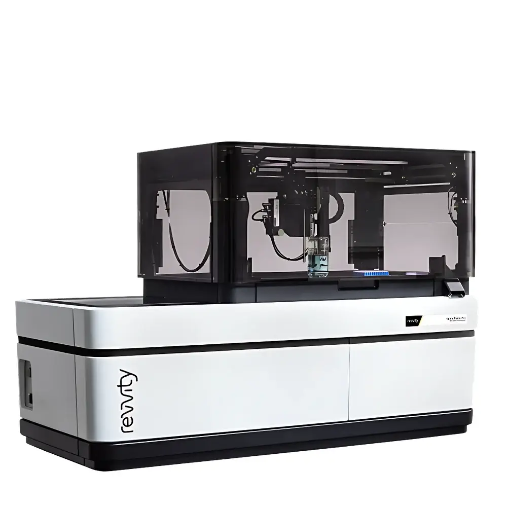
Revvity OPERA PHENIX PLUS High-Content Screening System
| Brand | Revvity |
|---|---|
| Origin | United Kingdom |
| Manufacturer | Revvity, Inc. |
| Type | Imported Instrument |
| Model | OPERA PHENIX PLUS |
| Temperature Control Range | 37–42 °C (±1 °C) |
| CO₂ Control Range | 1–10% (±0.5%) |
| Excitation Lasers | 405 nm, 488 nm, 561 nm, 640 nm (solid-state) |
| Brightfield Illumination | Near-infrared LED |
| Objective Type | High-NA water-immersion objectives |
| Imaging Throughput | ≥100,000 images per day |
| Detection | Up to four synchronized 16-bit sCMOS cameras |
| Confocal Technology | Dual Nipkow disk scanning |
| Software Platform | Acapella (with modular analysis workflows), optional Columbus cloud server and TIBCO Spotfire for advanced visualization |
Overview
The Revvity OPERA PHENIX PLUS High-Content Screening (HCS) System is an automated, laser-scanning confocal imaging platform engineered for quantitative, multi-parametric phenotypic analysis of live and fixed cells in microplate formats—from standard 96-well to high-density 1536-well and nanowell arrays. Built upon dual Nipkow spinning-disk confocal architecture, it delivers optical sectioning with minimal phototoxicity and high temporal resolution—critical for longitudinal live-cell assays. Unlike widefield systems, the dual-disk design enables simultaneous excitation and emission path optimization, significantly reducing crosstalk while preserving signal-to-noise ratio across multiplexed fluorescent channels. The system integrates tightly controlled environmental regulation (temperature ±1 °C, CO₂ ±0.5%), making it suitable for extended kinetic studies under physiologically relevant conditions. Its core application domain spans target identification, lead optimization, mechanism-of-action profiling, and cytotoxicity assessment in drug discovery pipelines compliant with GLP and early-stage GMP practices.
Key Features
- Four synchronized 16-bit scientific CMOS cameras enabling concurrent acquisition of fluorescence (up to four channels) and brightfield or Digital Phase Contrast (DPC) images without mechanical filter wheel delays.
- Dual Nipkow disk confocal optics with microlens-enhanced light throughput—optimized for high-speed z-stack acquisition at subcellular resolution (lateral resolution ≤250 nm, axial resolution ≤600 nm under standard conditions).
- Four solid-state lasers (405 nm, 488 nm, 561 nm, 640 nm) with precise intensity control and near-infrared LED for label-free brightfield illumination—supporting both endogenous contrast and multicolor immunofluorescence.
- High numerical aperture (NA) water-immersion objectives designed for deep-tissue penetration and uniform illumination across heterogeneous 3D cell models, including spheroids and organoids.
- Integrated environmental chamber maintaining temperature (37–42 °C) and CO₂ (1–10%) with real-time feedback monitoring—validated per ISO 13485-aligned calibration protocols.
- Modular software architecture supporting FDA 21 CFR Part 11-compliant audit trails, electronic signatures, and version-controlled analysis workflows.
Sample Compatibility & Compliance
The OPERA PHENIX PLUS accommodates a broad range of sample formats, including adherent and suspension cultures, primary cells, iPSC-derived lineages, co-cultures, and 3D microtissues in polymeric or hydrogel-based scaffolds. It supports ANSI/SLAS-standard microplates (96-, 384-, 1536-well) and custom nanowell arrays with automated focus map generation. All hardware and firmware comply with IEC 61000-6-2 (EMC immunity) and IEC 61010-1 (safety for laboratory equipment). Image acquisition and analysis modules are validated against ASTM E2925-21 (standard guide for HCS assay development) and support alignment with USP , ISO/IEC 17025, and internal QC/QA SOPs required for regulated bioassay environments.
Software & Data Management
Acapella software serves as the native acquisition and analysis engine—featuring drag-and-drop workflow builder, batch processing, and over 200 pre-validated analysis modules (e.g., nuclear translocation, mitochondrial fragmentation, neurite outgrowth, phagocytosis scoring). Data export conforms to MIACA (Minimum Information About a Cellular Assay) standards. Optional Columbus cloud-based image storage provides encrypted, audit-ready archiving with role-based access control and DICOM-SR compatibility. TIBCO Spotfire integration enables statistical modeling, heat map clustering, and interactive dashboarding for cross-assay meta-analysis—fully traceable from raw image to final report.
Applications
- Phenotypic screening for oncology, neurodegeneration, and metabolic disease targets using complex cellular readouts (e.g., autophagy flux, DNA damage response, synaptic density).
- Functional toxicology: real-time assessment of mitochondrial membrane potential, lysosomal pH, calcium oscillations, and caspase activation in primary hepatocytes or cardiomyocytes.
- 3D model characterization: quantification of proliferation gradients, hypoxia markers, and extracellular matrix remodeling in tumor spheroids and vascularized organoids.
- CRISPR/Cas9 validation: high-content confirmation of gene editing efficiency via multiplexed protein knockdown and morphological phenotype correlation.
- Immuno-oncology assays: spatial analysis of immune synapse formation, T-cell infiltration, and PD-L1 expression heterogeneity in co-culture systems.
FAQ
What regulatory standards does the OPERA PHENIX PLUS support for use in GxP environments?
It supports 21 CFR Part 11 compliance through Acapella’s electronic signature framework, audit trail logging, and data integrity controls—including immutable raw image storage and versioned analysis scripts.
Can the system be integrated into existing robotic automation platforms?
Yes—it features SLAS-compliant deck mapping, RS-232/Ethernet API, and native drivers for Revvity Cell::explorer, as well as third-party integrators including Hamilton STAR, Tecan Freedom EVO, and PerkinElmer JANUS.
Is z-stack acquisition supported for thick samples such as organoids?
Yes—automated autofocus, motorized objective nosepiece, and adaptive focus correction enable robust z-series capture up to 200 µm depth with consistent signal fidelity across layers.
How is photobleaching minimized during long-term live-cell imaging?
Through low-intensity laser exposure enabled by high-quantum-efficiency sCMOS sensors, pulsed illumination mode, and dynamic exposure adjustment based on real-time histogram feedback.
What file formats are natively supported for downstream analysis?
OME-TIFF (with embedded metadata), HDF5, and proprietary .nd2 formats; all export paths include standardized OME-XML annotation for FAIR data principles compliance.


