RWD nVista HD Miniaturized In Vivo Calcium Imaging System
| Brand | RWD |
|---|---|
| Origin | Guangdong, China |
| Manufacturer Type | Original Equipment Manufacturer (OEM) |
| Country of Origin | China |
| Model | nVista HD |
| Pricing | Available Upon Request |
Overview
The RWD nVista HD is a miniaturized, end-to-end in vivo calcium imaging platform engineered for high-resolution, large-scale neural circuit dynamics recording in freely behaving mice. It operates on the principle of fluorescence microscopy using genetically encoded calcium indicators (e.g., GCaMP3, GCaMP6, jGCaMP7), where transient increases in intracellular Ca²⁺ concentration—correlating with neuronal spiking activity—are transduced into quantifiable fluorescent signals. Unlike conventional benchtop two-photon or widefield microscopes requiring head fixation or anesthesia, the nVista HD integrates all optical, illumination, and acquisition components into a single lightweight (<2.5 g), head-mounted module. This enables chronic, cell-resolved functional imaging across cortical and subcortical regions—including hippocampus, amygdala, and striatum—during naturalistic behaviors such as spatial navigation, social interaction, operant conditioning, and motor learning. Its design adheres to principles of minimal perturbation, mechanical stability, and optical fidelity, supporting longitudinal studies under ethologically relevant conditions.
Key Features
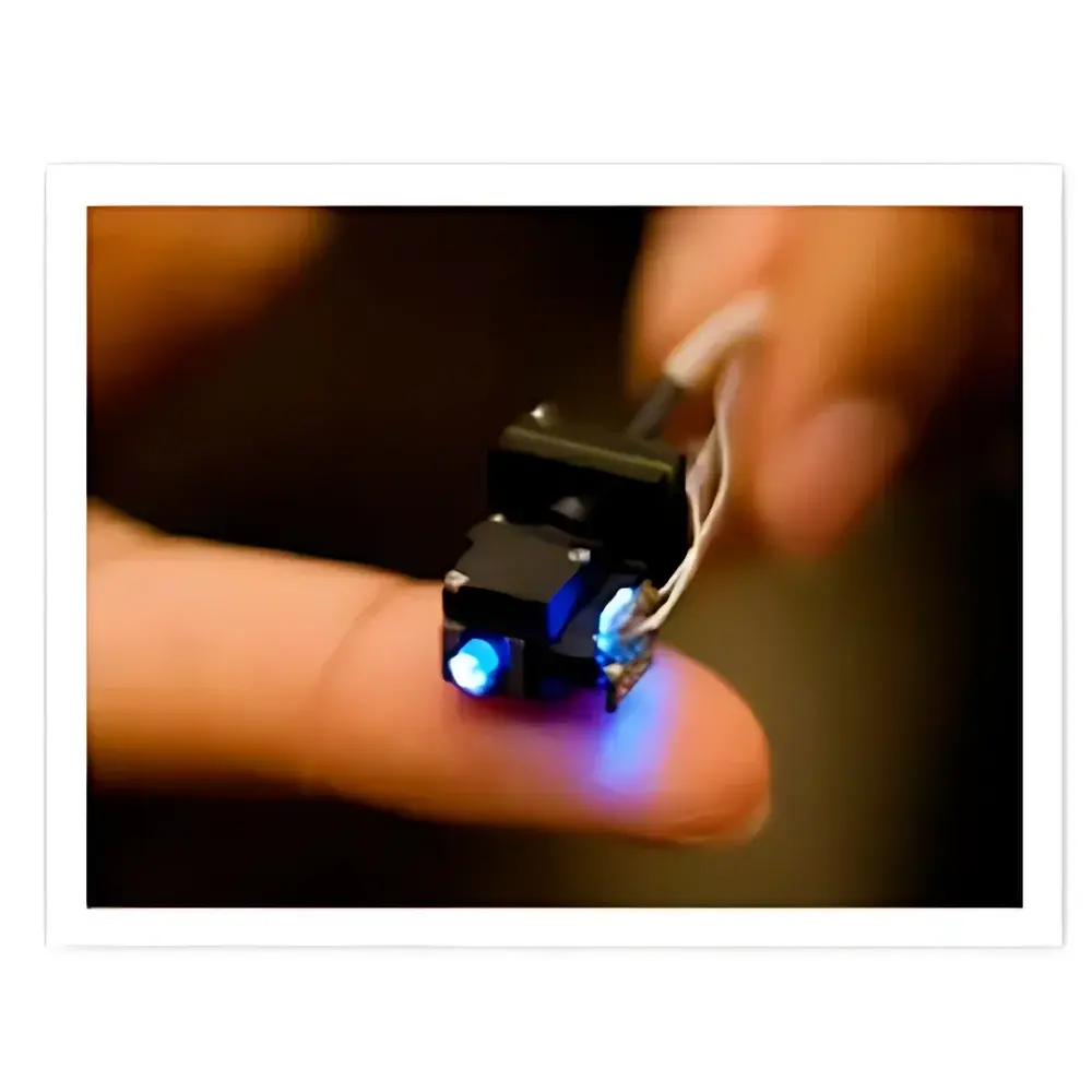
- Miniaturized integrated fluorescence microscope: All optical elements—including excitation LED, emission filter, objective lens, and CMOS sensor—are housed within a fingertip-sized (Ø 8.5 mm × 12 mm), biocompatible titanium housing.
- High-density neuronal recording: Simultaneous functional imaging of >1,000 individual neurons per field-of-view at cellular resolution (lateral resolution ≤ 5 µm, axial resolution ≤ 25 µm).
- Freely behaving paradigm compatibility: Fully USB-powered data acquisition module enables real-time streaming without external rack-mounted hardware or vibration-isolated tables.
- Surgical integration support: Includes Pro View™ lens probe set (3 interchangeable probes: 0.5 mm, 1.0 mm, and 1.5 mm working distance), stereotaxic-compatible lens holder, and craniotomy alignment guide for precise implantation.
- Indicator-agnostic optical path: Optimized for GFP-family fluorophores (excitation 450–490 nm, emission 500–550 nm), compatible with GCaMP variants, synthetic dyes (e.g., Oregon Green BAPTA-1), and red-shifted indicators (e.g., jRGECO1a) via optional filter swaps.
Sample Compatibility & Compliance
The nVista HD supports chronic implantation in adult C57BL/6, Thy1-GCaMP6s, Ai95, and other transgenic mouse lines. Surgical protocols align with NIH Guide for the Care and Use of Laboratory Animals and AAALAC International standards. The system’s modular design facilitates compliance with GLP-aligned experimental workflows: raw image streams are timestamped, metadata-tagged (animal ID, session date, probe ID), and stored in vendor-neutral HDF5 format—enabling traceability for regulatory submissions. While not FDA-cleared for clinical use, its architecture meets foundational requirements for preclinical neuropharmacology and behavioral phenotyping studies referenced in ICH S5(R3) and OECD TG 426 guidelines.
Software & Data Management
Acquisition and preprocessing are managed via nVista Control Suite—a cross-platform application (Windows/macOS) supporting real-time preview, focus adjustment, exposure control, and ROI-based signal extraction. The software exports time-series fluorescence traces (ΔF/F₀) in MATLAB-compatible .mat files and supports batch processing through Python API (nVistaPy). All acquisition logs include audit trail metadata (user login, parameter changes, hardware calibration timestamps), satisfying basic 21 CFR Part 11 documentation requirements for research integrity. Exported datasets are structured to integrate with open-source analysis pipelines including CaImAn, Suite2p, and SIMA.
Applications
- Longitudinal circuit plasticity: Tracking ensemble-level reorganization during learning across days or weeks.
- Deep-brain structure interrogation: Targeted imaging of ventral tegmental area (VTA), nucleus accumbens (NAc), or basolateral amygdala (BLA) using custom cannula-guided probe insertion.
- Behavioral state decoding: Correlating calcium dynamics with video-synchronized ethograms (e.g., motion onset, freezing, sniffing) via TTL-triggered synchronization.
- Pharmacological intervention studies: Monitoring acute or chronic drug effects on network synchrony, event rate, or inter-regional coupling metrics.
- Cross-lab reproducibility: Standardized probe geometry, excitation intensity, and frame rate (up to 30 Hz) facilitate multi-center comparative studies.
FAQ
Is the nVista HD compatible with head-fixed preparations?
Yes—while optimized for freely moving paradigms, the system can be adapted for head-fixed imaging by mounting the data acquisition module on a stable stage and using a lightweight commutator.
What is the maximum imaging depth achievable with standard probes?
With the 1.5 mm working distance Pro View™ probe and cleared-skull preparation, effective imaging depth reaches ~300 µm below pia; deeper targets require thinned-skull or cranial window surgery.
Does the system support dual-color or ratiometric calcium imaging?
Not natively—single-band excitation/emission is standard. Dual-channel capability requires third-party filter cube integration and external synchronization logic.
How is thermal management handled during extended recording sessions?
The LED driver implements pulse-width modulation (PWM) with duty cycling to limit average power dissipation; surface temperature rise remains <1.2°C above ambient over 60-minute continuous operation.
Can raw image data be exported without proprietary compression?
Yes—uncompressed 16-bit TIFF stacks and time-stamped HDF5 files are available directly from the acquisition buffer, enabling full transparency in downstream analysis.


