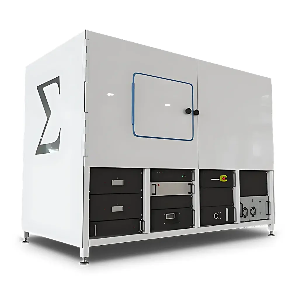
Sigray TriLambdaXRM-30 Nanoscale X-ray Microscope CT System
| Brand | Sigray |
|---|---|
| Origin | Shanghai, China |
| Model | TriLambdaXRM-30 |
| Spatial Resolution | 35 nm (at 1.7 keV) |
| X-ray Energy Range | 1.7–8.04 keV |
| Max. Number of Selectable Anode Targets | 3 (e.g., Si, Cr, Fe, Cu) |
| Source Type | Patented Multi-Target Reflective X-ray Source with Diamond Heat Sink |
| Optical Configuration | Triple Parabolic X-ray Optics + Fresnel Zone Plate Objective Lens |
| Phase Ring | Zernike Phase-Shifting Ring |
| Reconstructed Volume Resolution | Down to 65 nm voxel (LFOV mode) |
| Field of View (LFOV) | 65 µm diameter |
| FOV (HR mode) | ~10–20 µm |
| Reconstruction Software | GigaRecon (iterative CBCT reconstruction, <45 s for 2048×2048×2200 dataset) |
| Acquisition Software | Sigray3D with AI-assisted AutoPilot & Sample Handling Robot (SHR) integration |
| Control Architecture | Dual-Workstation (Windows GUI + Linux real-time EPICS-based control) |
| Dimensions | 2.3 m (L) × 1.3 m (W) × 1.5 m (H) |
| Compliance | Designed for GLP/GMP-aligned workflows |
Overview
The Sigray TriLambdaXRM-30 is a laboratory-scale nanoscale X-ray microscope (oXRM) engineered for true 3D non-destructive tomographic imaging at spatial resolutions down to 35 nm. Unlike lensless X-ray microtomography systems—such as those based on coherent diffraction imaging or phase retrieval—the TriLambdaXRM-30 employs a proprietary optical architecture integrating dual-parabolic reflective X-ray optics and Fresnel zone plate objective lenses. This hybrid focusing approach enables high-brightness, high-coherence illumination while maintaining quantitative absorption contrast across a broad energy range (1.7–8.04 keV). The system operates on the principle of absorption- and phase-enhanced X-ray computed tomography (CT), where differential attenuation and refraction at nanoscale interfaces are resolved through high-fidelity projection acquisition and iterative reconstruction. Its design targets applications requiring metrological-grade volumetric characterization of heterogeneous materials—including battery electrodes, catalyst architectures, semiconductor interconnects, and biological mineralized tissues—where sub-100 nm structural fidelity is essential for functional correlation.
Key Features
- Sub-40 nm isotropic spatial resolution (35 nm at 1.7 keV), achieved via Sigray’s patented triple parabolic X-ray optics coupled with high-efficiency Fresnel zone plate objectives.
- Tri-Lambda multi-energy source: Up to three selectable anode targets (e.g., Si, Cr, Fe, Cu) integrated into a single diamond-cooled reflective X-ray source, enabling rapid in situ switching between characteristic X-ray lines optimized for material-specific contrast.
- Zernike phase-shifting ring for enhanced edge contrast in low-absorption samples, improving segmentation accuracy for polymer matrices, soft biological specimens, and low-Z composites.
- Dual-energy nano-CT capability: Simultaneous or sequential acquisition at two discrete energies allows decomposition of linear attenuation coefficients into density- and atomic number-dependent components—critical for quantifying multi-phase alloys, mineral distributions, or nanoparticle dispersion.
- Dual-workstation architecture: Windows-based Sigray3D GUI for intuitive sample alignment, acquisition planning, and real-time preview; Linux-based EPICS-controlled subsystem for deterministic motion, detector synchronization, and 24/7 unattended operation.
- Integrated Sample Handling Robot (SHR): Enables automated batch processing of up to 12 standard pin-mount samples, supporting overnight and weekend experiments without manual intervention.
Sample Compatibility & Compliance
The TriLambdaXRM-30 accommodates samples ranging from 100 µm to 5 mm in diameter, with optimal performance observed for specimens ≤2 mm in thickness at high-resolution modes. It supports both conductive and non-conductive materials—including ceramics, metal alloys, polymers, geological thin sections, freeze-dried cells, and MEMS devices—without mandatory metallization or staining. The system adheres to international standards governing quantitative micro-CT: reconstructed volumes comply with ASTM E1441 (Standard Guide for Computed Tomography), ISO 12795 (Micro-CT terminology), and support traceable calibration via NIST-traceable step wedges and sphere phantoms. Data provenance is preserved through embedded metadata (acquisition parameters, source settings, geometry corrections), and the software stack supports FDA 21 CFR Part 11-compliant audit trails when deployed in regulated environments (e.g., medical device R&D, pharmaceutical formulation analysis).
Software & Data Management
Sigray3D provides a unified graphical interface for sample navigation, tilt-series definition, exposure optimization, and live projection monitoring. Its AI-driven AutoPilot module recommends optimal kVp, target selection, exposure time, and rotation step based on preliminary scout scans and user-defined objectives (e.g., “maximize contrast-to-noise ratio” or “minimize total dose”). GigaRecon delivers commercial-grade iterative reconstruction using ordered-subset expectation maximization (OSEM) and compressed sensing regularization, achieving artifact suppression and noise resilience unattainable with conventional FDK algorithms. All reconstructions output OME-TIFF stacks compliant with the Bio-Formats ecosystem and support direct import into Avizo, Dragonfly, and MATLAB. Raw projections and metadata are archived in HDF5 format, ensuring long-term readability and FAIR (Findable, Accessible, Interoperable, Reusable) data principles.
Applications
- Battery research: Quantification of SEI layer thickness, pore tortuosity, and Li dendrite morphology in cycled anodes at <50 nm resolution.
- Advanced manufacturing: Defect mapping in additively manufactured Ti-6Al-4V lattice structures, including microporosity and unmelted powder detection.
- Catalysis: 3D visualization of Pt nanoparticle distribution within mesoporous Al₂O₃ supports and pore-blocking analysis under operando-relevant conditions.
- Geosciences: Mineral phase identification and fluid inclusion geometry in shale matrix samples via dual-energy decomposition.
- Life sciences: Label-free 3D cytoarchitecture of calcified osteocytes in bone biopsies, preserving native hydration states through cryo-compatible sample stages (optional).
FAQ
What distinguishes TriLambdaXRM-30 from lensless X-ray microscopes like EclipseXRM?
The TriLambdaXRM-30 uses refractive/reflective X-ray optics to achieve ~10× higher spatial resolution than comparable lensless systems, enabling direct imaging of features below 50 nm without reliance on computationally intensive phase retrieval.
Can the system perform in situ or operando experiments?
Yes—mechanical stages support heating (up to 800 °C), cooling (down to −180 °C), and tensile loading (500 N max); custom environmental chambers integrate seamlessly with Sigray3D control logic.
Is GigaRecon compatible with third-party reconstruction pipelines?
GigaRecon exports raw sinograms and geometry files in standardized formats (e.g., HDF5, TIF stack with XML metadata), allowing interoperability with TomoPy, ASTRA Toolbox, or custom CUDA-accelerated solvers.
How is beamtime allocated for multi-user facilities?
The EPICS-based control layer supports role-based access control (RBAC), queue management, and reservation scheduling via RESTful API—enabling integration with facility-wide LIMS platforms.
Does the system meet regulatory requirements for quality-controlled environments?
Full 21 CFR Part 11 compliance is available via optional validation packages, including IQ/OQ documentation, electronic signature modules, and configurable audit trail retention policies aligned with ISO 13485 and ICH-GCP guidelines.


