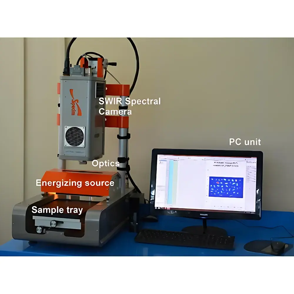
Specim sisuCHEMA Hyperspectral Imaging Analysis System
| Brand | Specim |
|---|---|
| Origin | Finland |
| Model | sisuCHEMA |
| Imaging Principle | Push-broom |
| Imaging Optics | Binary Optical Elements |
| Operating Environment | Ground-based |
| Spectral Ranges | 400–1000 nm (VNIR), 900–1700 nm (NIR), 1000–2500 nm (SWIR) |
| Spectral Resolution (FWHM) | 2.8 nm, 6 nm, 10 nm |
| Spatial Resolution | 38–152 µm (VNIR), 30–600 µm (NIR), 24–600 µm (SWIR) |
| Field of View (TFOV) | 38° |
| Instantaneous Field of View (IFOV) | 320 px, 384 px, 1312 px |
| Frame Rate | 670 fps |
Overview
The Specim sisuCHEMA Hyperspectral Imaging Analysis System is a turnkey laboratory-grade push-broom hyperspectral imaging workstation engineered for quantitative chemical mapping and spectral phenotyping across life sciences, geosciences, environmental monitoring, and industrial quality control. It integrates three interchangeable high-performance imaging spectrometers—covering the visible-near-infrared (VNIR: 400–1000 nm), near-infrared (NIR: 900–1700 nm), and short-wave infrared (SWIR: 1000–2500 nm) spectral domains—each optimized for distinct molecular absorption features (e.g., C–H, O–H, N–H overtones and combinations). The system employs binary optical elements to achieve diffraction-limited performance with minimal chromatic aberration, enabling high-fidelity spectral-spatial data acquisition under controlled illumination using Specim’s calibrated linear scattering light source. Designed for ground-based operation, it supports non-destructive, contactless analysis of solid or semi-solid samples up to 200 × 300 × 45 mm in dimension, with spatial sampling down to 30 µm for sub-millimeter specimens. All spectrometers are factory-calibrated for radiometric, spectral, and geometric accuracy; each scan includes automated reference-based calibration using integrated white/dark references to ensure traceable, reproducible measurements compliant with ISO/IEC 17025 requirements for analytical instrument validation.
Key Features
- Modular triple-band architecture: Interchangeable VNIR, NIR, and SWIR imaging spectrometers mounted on a shared precision motorized scanning stage.
- High-speed push-broom acquisition: Up to 670 full-frame spectra per second, enabling rapid mapping of heterogeneous samples without motion artifacts.
- Calibrated illumination: Specim linear scattering light source provides uniform, stable irradiance across the field of view, minimizing specular reflection and shadowing effects.
- Pixel-accurate spatial registration: Hardware-synchronized camera-spectrometer triggering ensures sub-pixel alignment between spectral bands and spatial coordinates.
- Robust mechanical design: Aluminum alloy frame with vibration-damped optical bench, suitable for integration into GLP-compliant laboratories or mobile analytical carts.
- BIL-format output: Native band-interleaved-by-line data export compatible with ENVI, Python (scikit-image, spectral), MATLAB, and commercial chemometric platforms.
Sample Compatibility & Compliance
The sisuCHEMA accommodates flat, planar, or low-relief samples placed on a motorized XYZ translation stage. Sample height tolerance is ±2 mm around the focal plane, supporting analysis of seeds, plant leaves, root segments, pharmaceutical tablets, mineral thin sections, polymer fragments, and food products. For biological tissues, no cryo-fixation or sectioning is required—samples remain intact throughout acquisition. The system complies with IEC 61000-6-3 (EMC emissions) and IEC 61000-6-2 (immunity), and its software architecture supports audit trails and electronic signatures per FDA 21 CFR Part 11 when deployed with validated LIMS integration. Data integrity is ensured via checksummed BIL files, timestamped metadata (including lamp intensity, ambient temperature, and stage position), and optional hardware-locked license enforcement.
Software & Data Management
sisuMAP software provides end-to-end workflow control—from instrument configuration and real-time preview to spectral library matching, multivariate image analysis (MIA), and false-color composition. Built-in tools include PCA, MCR-ALS, PLS-DA, and spectral angle mapper (SAM) algorithms for unsupervised and supervised classification. Users can generate quantitative concentration maps (e.g., chlorophyll-a, starch, lignin, moisture content) by applying pre-trained or user-defined regression models. All processing steps are scriptable via Python API (sisuPy), enabling reproducible batch analysis and integration into automated QA/QC pipelines. Raw and processed datasets are stored with embedded EXIF-like headers containing spectral calibration coefficients, exposure parameters, and geometric correction matrices—facilitating FAIR (Findable, Accessible, Interoperable, Reusable) data management per ISO 8000-100 standards.
Applications
- Plant Phenotyping: Quantification of pigment distribution, water status, nitrogen content, and disease biomarkers (e.g., anthracnose in cotton, Fusarium spp. colonization) at tissue-level resolution.
- Pharmaceutical & Food Science: Detection of counterfeit APIs, excipient homogeneity assessment, mycotoxin screening, and microplastic identification (PS/PE/PP discrimination in marine debris).
- Geoscience & Mining: Mineralogical mapping via diagnostic absorption features (e.g., Fe³⁺ crystal field transition at ~900 nm, Al–OH at 2200 nm), ore grade estimation, and hydrocarbon seep detection.
- Traditional Medicine: Non-invasive authentication of botanical origin and adulterant detection in herbal materials such as ginseng species.
- Environmental Monitoring: Soil organic carbon profiling, heavy metal stress indicators in vegetation, and phytoplankton community composition analysis from algal cultures.
FAQ
Is the sisuCHEMA suitable for live plant imaging?
Yes—its non-contact, non-destructive operation allows repeated longitudinal measurements on living specimens under ambient or controlled growth chamber lighting, provided motion is minimized during scan acquisition.
Can spectral libraries be imported from third-party sources?
Yes—sisuMAP accepts ASCII and ENVI-compatible spectral libraries (.sli, .asd, .spc), including USGS, ECOSTRESS, and JPL spectral databases, for spectral matching and endmember extraction.
What is the typical data size per scan?
A 320 × 320 pixel scan with 256 spectral bands yields approximately 25 MB in uncompressed BIL format; lossless compression reduces this by ~40% without spectral fidelity loss.
Does the system support remote operation and automation?
Yes—via Ethernet-connected API, the sisuCHEMA integrates with LabVIEW, Python, or custom scheduling software for unattended overnight scanning and trigger-based acquisition synchronized with environmental sensors.
Are calibration certificates provided with delivery?
Each unit ships with a traceable factory calibration report (NIST-traceable reference standards used), including wavelength accuracy (±0.2 nm), radiometric linearity (R² > 0.9999), and spatial distortion metrics (< 0.5% RMS).