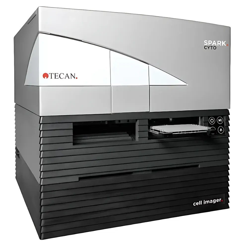
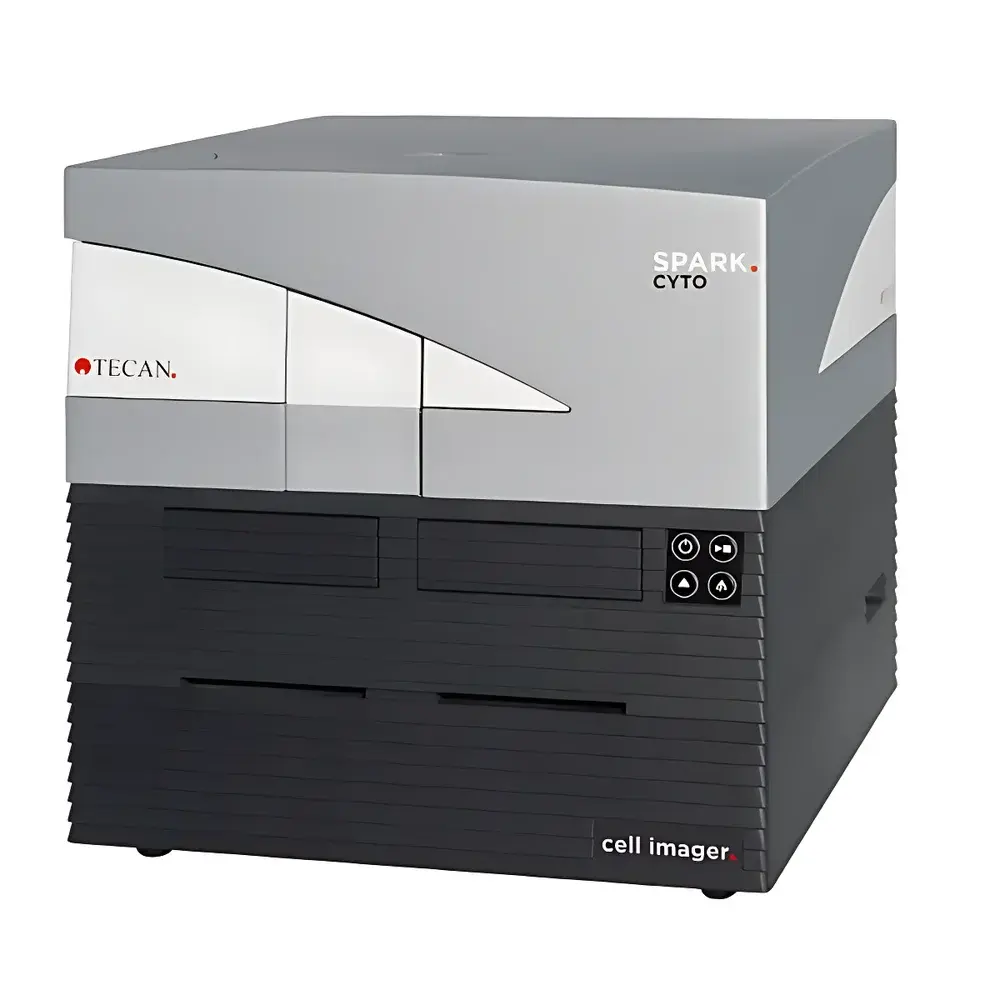
Tecan Spark Cyto Multimodal Live-Cell Imaging Microplate Reader
| Brand | Tecan |
|---|---|
| Origin | Austria |
| Model | Spark Cyto |
| Automation Level | Fully Automated |
| Detection Modes | Fluorescence Imaging, Brightfield, Multimode Plate Reading (Absorbance, Fluorescence Intensity, Luminescence, Time-Resolved Fluorescence, FRET) |
| Imaging Capabilities | Widefield Epifluorescence, Full-Well Capture per Field, Z-Stacking, Kinetic Time-Lapse, 2D & 3D Cell Culture Compatibility |
| Objectives | 2×, 4×, 10× (Motorized, Auto-Focus Enabled) |
| Fluorescence Channels | 4 Independent Filter Sets (Configurable) |
| Software Platform | SparkControl™ MAGNA with Real-Time Experiment Control (REC), GLP-Compliant Audit Trail, FDA 21 CFR Part 11 Ready |
| Throughput Options | Standalone or Integrated with Tecan Fluent/Genesis Liquid Handling Platforms and Incubation Modules (e.g., Gas-Controlled Multi-Plate Incubator) |
Overview
The Tecan Spark Cyto is a purpose-engineered multimodal microplate reader that integrates high-resolution widefield epifluorescence imaging with industry-standard multimode detection capabilities—including absorbance, fluorescence intensity, luminescence, time-resolved fluorescence (TRF), and Förster resonance energy transfer (FRET). Unlike conventional plate readers limited to well-averaged signal outputs, the Spark Cyto captures spatially resolved, single-cell–level morphological and functional data directly from 2D monolayers and 3D spheroid/organoid cultures in standard microplates (96-, 384-, and 1536-well formats). Its optical architecture is based on a high-quantum-efficiency sCMOS sensor coupled with motorized, parfocal objectives and precision Z-drive mechanics—enabling consistent focus maintenance across kinetic and Z-stack acquisitions. Designed for longitudinal live-cell experiments, the system maintains physiological relevance through integrated environmental control compatibility (CO₂, O₂, humidity, temperature) and real-time experiment control (REC) functionality, allowing dynamic parameter adjustment during acquisition without interrupting workflow continuity.
Key Features
- Full-well imaging in a single capture: Eliminates tiling artifacts and geometric distortion across 96- and 384-well plates using optimized widefield optics and calibrated pixel mapping.
- Triple-motorized objective turret (2×, 4×, 10×) with auto-focus and parfocal alignment—ensuring reproducible magnification switching and Z-position consistency across multi-day kinetics.
- Four independently configurable fluorescence channels with high-transmission bandpass filters and LED excitation sources—optimized for common fluorophores including DAPI, FITC, TRITC, Cy5, and Hoechst variants.
- Simultaneous multimode detection and image acquisition: Enables orthogonal validation—e.g., correlating caspase-3 activation (fluorescence assay) with nuclear condensation (Hoechst imaging) within the same well and timepoint.
- Real-Time Experiment Control (REC): Adjust exposure time, gain, Z-step interval, or incubation parameters mid-run via software interface—critical for adaptive response profiling in stress assays or drug washout studies.
- FDA 21 CFR Part 11–ready software architecture with electronic signatures, full audit trail, and role-based access control—supporting regulated environments under GLP/GMP frameworks.
Sample Compatibility & Compliance
The Spark Cyto supports diverse biological models, including adherent and suspension cell lines, primary neurons, iPSC-derived cardiomyocytes, co-cultures, and scaffold-free 3D spheroids up to 500 µm in diameter. Its large field-of-view optics and low-magnification objectives (2×, 4×) accommodate heterogeneous cell distributions while preserving cellular context—essential for phenotypic screening and toxicity assessment. The platform complies with ISO/IEC 17025 calibration traceability standards for photometric and fluorescence intensity measurements. All imaging and detection protocols are documented and exportable in MIAME/MIAPE-compliant metadata structures. Environmental integration modules meet ASTM E1985-22 specifications for CO₂ and temperature stability in long-term incubation applications.
Software & Data Management
SparkControl™ MAGNA provides an integrated environment for method development, acquisition, analysis, and reporting. Built-in image analysis modules support segmentation (cell/nucleus/membrane), confluence quantification, object counting, intensity distribution profiling, and motion tracking—without requiring third-party plugins. Data is stored in vendor-neutral HDF5 format with embedded metadata (acquisition parameters, instrument logs, user annotations). Batch processing pipelines support parallel analysis of multi-plate datasets, and RESTful API access enables integration with LIMS (e.g., LabVantage, STARLIMS) and ELN systems (e.g., Benchling, LabArchives). Audit trails record all user actions—including parameter edits, image reprocessing, and report generation—with immutable timestamps and operator IDs.
Applications
- High-content cytotoxicity screening: Quantify membrane integrity (propidium iodide), mitochondrial potential (TMRM), and apoptosis markers (Annexin V-FITC) in parallel with morphology.
- 3D tumor spheroid growth and invasion monitoring: Track volume changes, necrotic core formation, and edge migration over 7–14 days using brightfield + fluorescent viability dyes.
- Neurite outgrowth and network maturation analysis: Measure total neurite length, branching points, and synaptic puncta density in primary neuronal cultures.
- Stem cell differentiation kinetics: Correlate lineage-specific reporter expression (e.g., GFP-tagged SOX2 or OCT4) with morphological transitions in real time.
- Functional calcium imaging: Perform ratiometric (Fura-2) or single-wavelength (Fluo-4) kinetics in microplate format with sub-second temporal resolution.
- Label-free label retention assays: Combine phase contrast imaging with fluorescence readouts to assess transfection efficiency or CRISPR editing outcomes.
FAQ
Does Spark Cyto support confocal imaging?
No—Spark Cyto utilizes widefield epifluorescence optics optimized for speed, sensitivity, and uniform illumination across full wells. Confocal capability is not included; however, deconvolution algorithms are available in SparkControl™ MAGNA to enhance axial resolution in Z-stacks.
Can it image cells in opaque or black-walled plates?
Yes—brightfield and fluorescence imaging modes are compatible with standard tissue-culture-treated black, white, and clear-bottom microplates. Opaque plates require bottom-read configuration with appropriate objective working distance.
Is Z-stack acquisition automated and software-controlled?
Yes—Z-stacking is fully programmable with user-defined step size (0.1–10 µm), number of slices, and autofocus strategy (contrast-based or hardware-assisted). Stacks are acquired sequentially per well and aligned post-acquisition.
How does Spark Cyto ensure data integrity in regulated labs?
Through FDA 21 CFR Part 11–compliant features: electronic signatures, audit trail logging, data encryption at rest, and secure user authentication—all validated and documented per IQ/OQ/PQ protocols.
What liquid handling integrations are supported?
Native compatibility with Tecan Fluent and Genesis platforms; robotic arm interfaces follow ANSI/SLAS standards. Third-party integration is possible via RS-232, TCP/IP, or OPC UA protocols.


