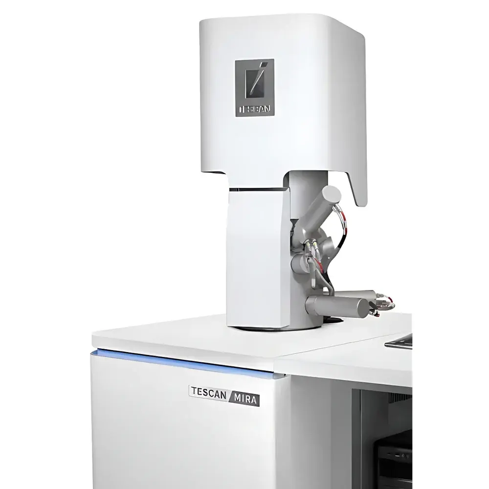
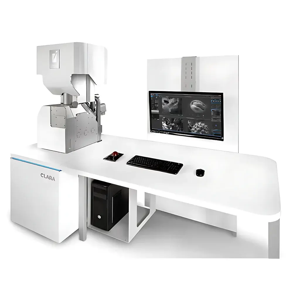
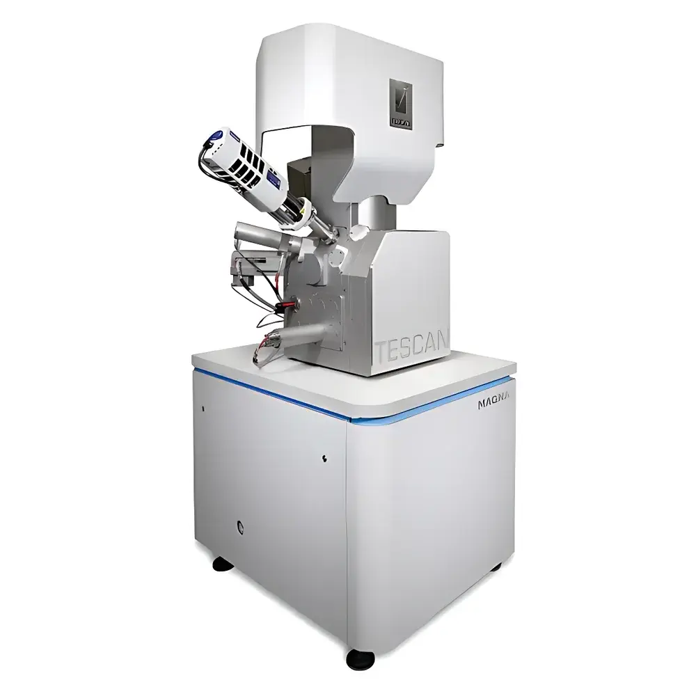
TESCAN MIRA/CLARA/MAGNA Series Field Emission Scanning Electron Microscopes
| Brand | TESCAN |
|---|---|
| Origin | Czech Republic |
| Model(s) | MIRA / CLARA / MAGNA |
| Instrument Type | Floor-standing FE-SEM |
| Electron Source | Schottky Field Emission Gun |
| Beam Technology | BrightBeam (CLARA), Triglav (MAGNA) |
| Primary Application Domains | Semiconductor Metrology, Advanced Materials Characterization, Life Sciences Imaging, Geoscience Microanalysis |
Overview
The TESCAN MIRA, CLARA, and MAGNA series represent a family of high-performance field emission scanning electron microscopes (FE-SEMs) engineered for nanoscale surface imaging, compositional analysis, and structural characterization across diverse scientific and industrial disciplines. Operating on the fundamental principle of raster-scanned electron beam interaction with solid specimens, these instruments generate secondary electrons (SE), backscattered electrons (BSE), and characteristic X-rays to deliver quantitative topographic, crystallographic, and elemental data. Unlike thermionic sources, the integrated Schottky field emission electron gun provides exceptional brightness, energy stability, and long-term emission reproducibility—enabling sub-nanometer resolution at low accelerating voltages (0.1–30 kV) while minimizing beam-induced damage. Each platform is purpose-built: MIRA delivers robust routine high-resolution imaging; CLARA introduces the patented BrightBeam electron column—a lens-free, ultra-low magnetic field design that eliminates image distortion on magnetic and beam-sensitive samples; MAGNA integrates the Triglav electron optical system, combining aberration-corrected optics with multi-detector signal synergy to maximize surface sensitivity and contrast fidelity for insulating or electron-beam-labile materials.
Key Features
- Schottky field emission source with >2,500-hour operational lifetime and <0.5 eV energy spread for stable, high-current probe formation
- Multi-mode imaging: In-lens SE, through-the-lens BSE, low-voltage SE, and immersion mode for enhanced surface detail
- Integrated beam deceleration (LVD) enabling high-resolution imaging at landing energies as low as 50 eV—critical for uncoated polymers, biological tissues, and thin-film devices
- Modular detector architecture: Everhart-Thornley SE detector, solid-state BSE detector, STEM-in-SEM detector, and optional energy-dispersive X-ray spectrometer (EDS) with silicon drift detector (SDD)
- Motorized 5-axis eucentric stage with ±70° tilt, 360° rotation, and precise Z-height control for comprehensive 3D reconstruction and cross-sectional analysis
- Vacuum system with differential pumping stages maintaining <1×10⁻⁷ Pa in the specimen chamber during operation—even with EDS or EBSD integration
Sample Compatibility & Compliance
These FE-SEMs accommodate a broad range of conductive and non-conductive specimens—including bulk metals, sintered ceramics, semiconductor wafers, freeze-dried biological sections, geological thin sections, and fiber-reinforced composites—without mandatory metal coating when operated in low-voltage or charge-compensation modes. The CLARA’s zero-magnetic-field column permits direct imaging of ferromagnetic materials (e.g., Fe, Ni, Co alloys, permanent magnets) without demagnetization or edge artifacts. MAGNA’s Triglav column enables stable imaging of beam-sensitive organics (e.g., MOFs, pharmaceutical crystals, lipid bilayers) at <1 kV with real-time charge neutralization. All systems comply with IEC 61000-6-3 (EMC), ISO 14644-1 Class 5 cleanroom compatibility for wafer metrology applications, and support GLP/GMP audit trails via optional software modules aligned with FDA 21 CFR Part 11 requirements.
Software & Data Management
Acquisition and analysis are managed through TESCAN’s Unified Platform (UP) software—a modular, scriptable environment supporting automated workflows, batch acquisition, and AI-assisted feature recognition. UP includes real-time drift correction, auto-focus/astigmatism routines, and integrated EDS mapping quantification compliant with ISO 14782 and ASTM E1508 standards. Data formats adhere to open standards (TIFF, HDF5, EMF) for interoperability with third-party analysis tools (e.g., DigitalMicrograph, ImageJ, MATLAB). Raw image metadata embeds full acquisition parameters—including kV, working distance, dwell time, detector configuration, and vacuum status—for traceable reporting in regulated environments.
Applications
- Semiconductor & Microelectronics: Critical dimension measurement of TSVs, Cu interconnects, and BEOL structures; failure analysis of solder joints and die attach; contamination identification via EDS correlation
- Advanced Materials: Grain boundary analysis in superalloys, phase distribution in thermal barrier coatings, porosity quantification in additively manufactured Ti-6Al-4V, and dispersion homogeneity in nanocomposites
- Life Sciences: Ultrastructural imaging of cryo-fractured cell membranes, collagen fibril orientation in decellularized scaffolds, nanoparticle uptake in macrophages, and correlative light-electron microscopy (CLEM) registration
- Geoscience & Environmental: Mineral phase identification and textural classification in shale cores, microfossil morphology reconstruction, particulate matter source attribution (PM₂.₅), and leaching interface characterization in nuclear waste forms
FAQ
What is the typical resolution specification for the TESCAN MAGNA at 1 kV?
Resolution is sample- and condition-dependent; under optimal conditions (conductive sample, in-lens SE detection, 1 kV, WD = 3 mm), MAGNA achieves ≤1.0 nm resolution per ISO 19749.
Can the CLARA system image uncoated biological tissue without charging artifacts?
Yes—CLARA’s BrightBeam column enables stable low-kV (<1 kV) imaging with simultaneous beam deceleration and active charge compensation, routinely used for hydrated or resin-embedded tissue sections without sputter coating.
Is EBSD integration supported on all three platforms?
EBSD is fully compatible with MIRA and MAGNA via dedicated ports and synchronized acquisition; CLARA supports EBSD with optional beam path modification due to its unique column geometry.
How does TESCAN ensure long-term calibration stability for quantitative EDS analysis?
Each system includes factory-calibrated detector efficiency curves, reference standards for ZAF matrix correction, and software-based dead-time and pulse pile-up correction validated against NIST SRM 2100a.
Are remote operation and multi-user access supported?
Yes—UP software supports secure remote desktop access, role-based user permissions, and concurrent multi-client sessions with session logging for audit compliance.


