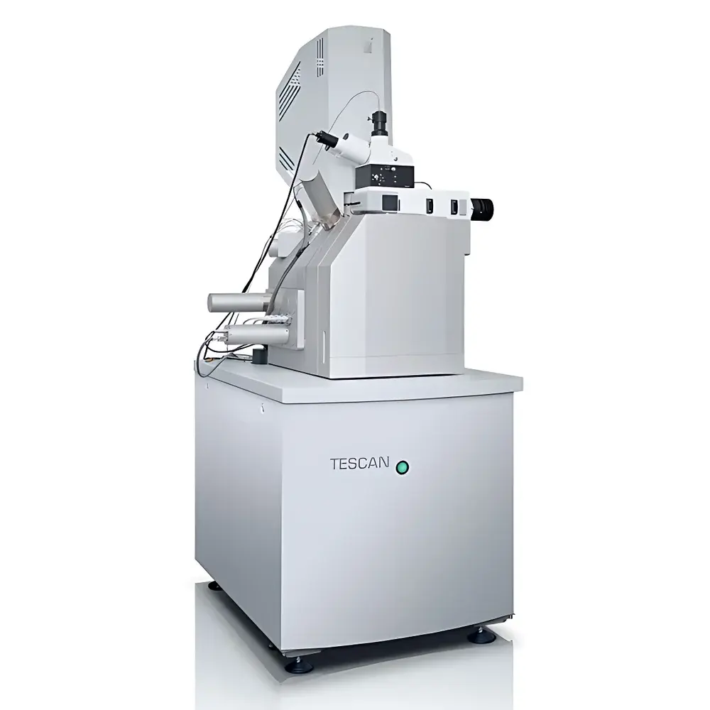
TESCAN RISE Microscope – Integrated Scanning Electron Microscope and Confocal Raman Imaging System
| Brand | TESCAN |
|---|---|
| Origin | Czech Republic |
| Manufacturer Type | Original Equipment Manufacturer (OEM) |
| Import Category | Imported Instrument |
| Model | RISE Microscope |
| Electron Gun Type | Cold Field Emission |
| Secondary Electron Image Resolution | 1.0 nm @ 15 kV |
| Accelerating Voltage Range | 0.2–30 kV |
| Backscattered Electron Image Resolution | 1.0 nm @ 15 kV |
| Confocal Raman Spatial Resolution | 360 nm (with 532 nm laser) |
| Optical Architecture | Parallel-Axis Co-Localized SEM–Raman Design |
| Maximum Sample Chamber Capacity | Compatible with TESCAN’s Ultra-Large Chamber Platforms |
| Detector Compatibility | Full Integration with BSE, CL, EDS, and Other In-Chamber Detectors |
Overview
The TESCAN RISE Microscope is a fully integrated scanning electron microscope (SEM) and confocal Raman imaging system engineered for correlative nanoscale structural and molecular characterization. Unlike conventional sequential or loosely coupled hybrid instruments, the RISE platform implements a true parallel-axis optical and electron-beam architecture—enabling simultaneous, spatially registered acquisition of high-resolution topographic, compositional, and vibrational spectroscopic data from the exact same micro/nanoscale region. Its core principle relies on precise co-localization of a cold field emission electron beam and a diffraction-limited, high-numerical-aperture (NA) confocal Raman excitation path within a single vacuum-compatible sample chamber. This design eliminates mechanical repositioning errors and ensures sub-micrometer registration fidelity between SEM morphology and Raman chemical maps—critical for analyzing heterogeneous materials such as mineral inclusions, polymer blends, carbon allotropes, pharmaceutical crystallites, and biological tissue sections.
Key Features
- Parallel-axis co-localized SEM–Raman architecture ensuring <100 nm spatial registration accuracy between electron and optical signals
- Cold field emission electron source delivering stable, high-brightness imaging at accelerating voltages from 0.2 to 30 kV
- Sub-nanometer secondary and backscattered electron resolution (1.0 nm @ 15 kV), compliant with ISO 16700:2016 for SEM performance verification
- Confocal Raman imaging with 360 nm lateral resolution using 532 nm excitation—surpassing classical diffraction limits via high-NA objective coupling and precision stage scanning
- Full compatibility with standard TESCAN in-chamber detectors: energy-dispersive X-ray spectrometer (EDS), cathodoluminescence (CL), backscattered electron (BSE), and electron backscatter diffraction (EBSD)
- Independent operational modes: SEM-only, Raman-only, and fully synchronized correlative acquisition
- X-Position function enabling overlay and alignment of Raman maps with external optical microscopy images, AFM topography, or EDS elemental distributions
- Automated point-line-area Raman mapping with real-time spectral fitting, baseline correction, and multivariate analysis (e.g., PCA, cluster analysis)
Sample Compatibility & Compliance
The RISE Microscope accommodates diverse sample types—including conductive and non-conductive solids, polished thin sections, bulk geological specimens, polymer films, battery electrode cross-sections, and fixed biological tissues—without requiring extensive pre-treatment. Conductive coating is optional and only applied when necessary for charge mitigation during high-current SEM imaging; Raman acquisition remains unaffected due to optical access through dedicated viewport windows. The system complies with IEC 61000-6-3 (EMC emissions) and IEC 61000-6-4 (industrial immunity), and its vacuum interlock and radiation shielding meet EU Machinery Directive 2006/42/EC requirements. For regulated environments, RISE supports audit-ready operation under GLP and GMP frameworks through optional software modules enabling 21 CFR Part 11-compliant electronic signatures, user access control, and full audit trail logging of all acquisition and processing parameters.
Software & Data Management
Acquisition and analysis are managed via TESCAN’s Unified Platform (UP) software suite, which provides a unified interface for both SEM control and Raman spectral processing. The platform supports vendor-neutral data formats (HDF5, SPE, JCAMP-DX) and includes built-in tools for spectral deconvolution, peak assignment against commercial libraries (e.g., RRUFF, ICDD PDF-4+), false-color mapping, and hyperspectral unmixing. All raw spectra, metadata (including beam parameters, laser power, integration time, stage coordinates), and processed maps are stored with FAIR (Findable, Accessible, Interoperable, Reusable) principles in mind. Optional integration with LabArchives ELN or Thermo Fisher SampleManager LIMS enables traceable workflow linkage from instrument acquisition to final report generation—essential for quality-controlled laboratories operating under ISO/IEC 17025 accreditation.
Applications
The RISE Microscope delivers unique analytical capabilities across multiple domains requiring simultaneous morphological and molecular insight. In geosciences, it identifies polymorphic mineral phases (e.g., quartz vs. coesite), maps fluid inclusion chemistry, and correlates crystallographic orientation (via EBSD) with local strain-induced Raman shifts. In advanced materials research, it resolves graphene layer stacking order, distinguishes sp²/sp³ carbon bonding in diamond-like carbon films, and quantifies crystallinity gradients in semi-crystalline polymers. In life sciences, it localizes drug distribution within tissue sections while preserving ultrastructural context, and characterizes calcification or amyloid deposition with subcellular spatial correlation. In forensics and gemology, it differentiates synthetic from natural gemstones based on trace-element-related Raman modes while documenting surface wear features at nanoscale resolution. These applications align with ASTM E2821–22 (Standard Guide for Correlative Microscopy) and support method validation per ISO 13847 for vibrational spectroscopy.
FAQ
Can the RISE system be retrofitted onto an existing TESCAN SEM?
Yes—the RISE configuration is available as a factory-integrated upgrade for compatible TESCAN MIRA and SCIMA series platforms, provided the chamber geometry and port layout meet mechanical and optical alignment specifications.
Is vacuum required during Raman acquisition?
No—Raman measurements are performed under ambient pressure through dedicated optical viewports; only SEM imaging requires high vacuum (≤1×10⁻⁴ Pa).
What laser wavelengths are supported?
Standard configurations include 532 nm and 785 nm lasers; optional 638 nm and UV (325 nm) sources are available for resonance-enhanced or deep-UV Raman applications.
Does the system support time-resolved or stimulated Raman scattering (SRS)?
No—the RISE platform is optimized for steady-state confocal Raman mapping; time-resolved or nonlinear variants require dedicated ultrafast laser systems outside this configuration.
How is calibration maintained across correlative modes?
Stage calibration is performed using NIST-traceable grid standards; optical-electron coordinate transformation is validated daily using dual-mode reference samples (e.g., Si grating with known pitch and characteristic Raman band).


