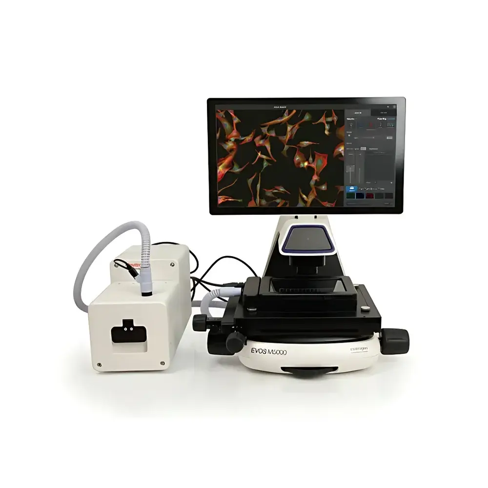
Thermo Fisher EVOS M5000 Live-Cell Fluorescence Imaging System
| Brand | Thermo Fisher |
|---|---|
| Origin | USA |
| Model | EVOS M5000 |
| Light Source | Adjustable LED Cube (Lifespan > 50,000 hrs per cube) |
| Imaging Modes | Fluorescence (4-channel simultaneous acquisition), Color Brightfield, Z-Stack, Time-Lapse, Maximum Intensity Projection (MIP), 3D Reconstruction |
| Focus Control | Motorized Auto-Focus with Software-Driven Z-Axis Control |
| Environmental Control | Optional Live-Cell Chamber (Temperature, Humidity, CO₂/O₂/N₂ Regulation) |
| Compliance | Designed for GLP/GMP-aligned workflows |
Overview
The Thermo Fisher EVOS M5000 Live-Cell Fluorescence Imaging System is an integrated upright/inverted hybrid platform engineered for high-fidelity, non-invasive optical sectioning and longitudinal monitoring of living biological specimens. Utilizing widefield fluorescence microscopy combined with precision motorized Z-axis actuation, the system captures volumetric image stacks via sequential focal plane acquisition—enabling quantitative analysis of spatially heterogeneous structures such as organoids, spheroids, neural networks, and multilayered tissue explants. Its optical architecture incorporates optimized excitation/emission filtering, high quantum efficiency CMOS imaging sensors, and diffraction-limited resolution across all four integrated fluorescence channels. Unlike confocal systems requiring laser scanning or point detection, the EVOS M5000 delivers rapid, low-phototoxicity imaging essential for maintaining physiological relevance during extended time-lapse experiments.
Key Features
- Motorized auto-focus algorithm with real-time contrast-based optimization, ensuring consistent in-focus acquisition across multi-well plates, chamber slides, and custom culture substrates.
- Z-Stack functionality with user-defined step size (0.1–10 µm increments) and up to 100 slice depth per stack, supporting both maximum intensity projection (MIP) and depth-coded 3D volume rendering.
- Four independently configurable fluorescence channels (e.g., DAPI, FITC, TRITC, Cy5), each driven by dedicated LED light cubes with spectral profiles matched to common fluorophores—minimizing cross-talk and enabling ratiometric quantification.
- Color brightfield mode utilizing white LED illumination and RGB sensor interpolation, providing morphological context without additional staining or phase contrast hardware.
- Modular live-cell environmental control unit (optional): maintains stable temperature (±0.2 °C from 20–40 °C), relative humidity (>95% RH), and gas composition (CO₂: 0–20%, O₂: 0–21%, N₂ balance) within sealed imaging chambers—validated for ≥72-hour uninterrupted assays.
- LED light cubes rated for >50,000 hours of operational life at nominal output, eliminating routine lamp replacement and reducing long-term maintenance overhead.
Sample Compatibility & Compliance
The EVOS M5000 accommodates standard microplate formats (6–384-well), glass-bottom dishes (e.g., MatTek, ibidi), flow chambers, and custom-engineered scaffolds used in 3D cell culture models. Its stage design supports both manual positioning and programmable coordinate mapping for high-content screening applications. All imaging metadata—including exposure time, gain, Z-step parameters, and environmental logs—are embedded in TIFF/OME-TIFF files, ensuring traceability required under ISO/IEC 17025, ASTM E2925, and USP guidelines. The system’s software architecture complies with FDA 21 CFR Part 11 through role-based access control, electronic signature support, and immutable audit trails for all acquisition and processing events—facilitating regulatory submissions in pharmaceutical development and academic GLP studies.
Software & Data Management
Acquisition and analysis are managed via the EVOS Imaging Software v3.x, a Windows-based application offering intuitive workflow scripting, batch processing, and export to open formats (TIFF, PNG, AVI, OME-Zarr). Built-in tools include background subtraction, flat-field correction, channel alignment, and pixel-intensity calibration using NIST-traceable reference standards. For advanced quantification, users may export ROI masks and intensity matrices to MATLAB, Python (via Bio-Formats API), or commercial platforms such as Imaris or Huygens. Raw data integrity is preserved through checksum validation and optional network-attached storage (NAS) integration with SMB/NFS protocols.
Applications
- Longitudinal tracking of neuronal outgrowth, synapse formation, and calcium dynamics in primary cortical cultures.
- Quantitative assessment of spheroid compaction, necrotic core development, and drug-induced apoptosis in tumor organoid models.
- Time-resolved visualization of endothelial barrier integrity under inflammatory cytokine challenge using dual-channel VE-cadherin/TJ protein labeling.
- High-content screening of CRISPR-edited iPSC-derived cardiomyocytes for contractile rhythm and mitochondrial membrane potential stability.
- Validation of 3D bioprinted constructs via structural co-localization of extracellular matrix components and cell viability markers.
FAQ
Is the EVOS M5000 compatible with third-party objective lenses?
Yes—the system accepts standard RMS-threaded objectives (4x–60x, NA 0.13–0.95), including phase contrast and apochromatic variants, provided mechanical clearance and back focal plane alignment are verified.
Can Z-Stack data be exported for external 3D reconstruction software?
Yes—all Z-series acquisitions are saved as multi-page TIFF or OME-TIFF files with embedded dimensional metadata, fully compatible with Fiji/ImageJ, Arivis Vision4D, and Bitplane Imaris.
Does the live-cell module support hypoxic conditions below 1% O₂?
Yes—the tri-gas controller enables O₂ setpoints from 0.1% to 21%, with nitrogen purge capability to achieve physiologically relevant hypoxia mimicking bone marrow or solid tumor microenvironments.
How is photobleaching minimized during multi-hour time-lapse experiments?
Through LED power modulation, adaptive exposure control, and hardware-triggered acquisition synchronized with stage movement—reducing cumulative photon dose by up to 60% compared to fixed-intensity protocols.
Is remote operation supported for core facility deployment?
Yes—secure RDP and web-based status monitoring (via optional Thermo Fisher Connect integration) allow off-site experiment initiation, progress tracking, and alert notifications without local workstation presence.


