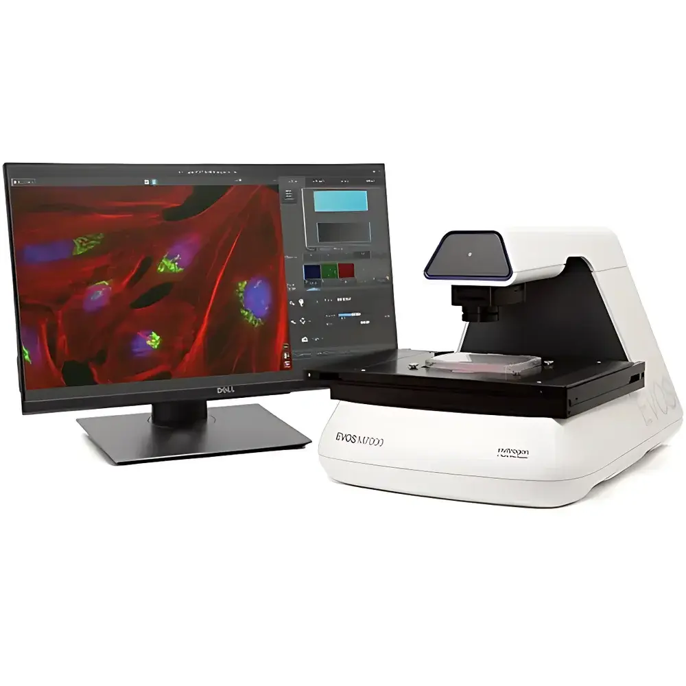
Thermo Fisher EVOS M7000 High-Content Cell Imaging System
| Brand | Thermo Fisher |
|---|---|
| Origin | USA |
| Model | AMF7000 |
| Optical System | Infinity-corrected, RMS-threaded objectives (45 mm parfocal distance) |
| Illumination | 4-position LED fluorescence cube turret + brightfield channel |
| Objectives | 5-position motorized turret |
| Cameras | Dual 3.2 MP CMOS (monochrome & color, 2048 × 1536, 3.45 µm pixel size) |
| Stage | Motorized X/Y scanning stage (120 mm × 80 mm travel, sub-micron resolution) |
| Focus | Automated autofocus with sub-micron precision |
| Display | 27-inch 4K monitor (3840 × 2160) |
| Software Platform | EVOS Acquisition & Analysis Suite (standard) |
| Compliance | Designed for GLP/GMP-aligned workflows |
Overview
The Thermo Fisher EVOS M7000 High-Content Cell Imaging System is an inverted, fully automated fluorescence and transmitted-light imaging platform engineered for reproducible, publication-grade cellular phenotyping in academic, pharmaceutical, and contract research laboratory environments. It operates on a robust optomechanical architecture grounded in infinity-corrected optics and digitally stabilized LED illumination—enabling quantitative multi-channel acquisition without mercury vapor lamp warm-up, spectral drift, or intensity decay. The system implements a dual-camera architecture (monochrome and color CMOS), synchronized with a 5-position objective turret and motorized X/Y/Z stage, to support concurrent acquisition of fluorescent, brightfield, phase contrast, and color brightfield modalities. Its design prioritizes experimental fidelity: hard-coated interference filters ensure >90% transmission efficiency and 50,000 hours; and the integrated environmental control interface enables seamless orchestration of time-lapse experiments under normoxic or hypoxic conditions via the optional EVOS Onstage Incubator. Unlike legacy widefield systems reliant on manual alignment or post-acquisition stitching, the EVOS M7000 embeds hardware-level synchronization between stage motion, focus adjustment, filter switching, and exposure timing—minimizing phototoxicity while maximizing spatial and temporal resolution consistency across multi-well plates, flasks, slides, and custom vessels.
Key Features
- Fully automated inverted imaging platform with synchronized control of 5-position objective turret, 4-channel LED fluorescence cubes, dual CMOS cameras (3.2 MP monochrome + 3.2 MP color), motorized X/Y stage (120 × 80 mm), and sub-micron Z-focus mechanism
- Smart LED illumination architecture featuring digitally tunable intensity, hard-coated filter sets, and >50,000-hour lifetime—eliminating lamp replacement cycles and enabling precise light dosing for live-cell assays
- Native support for multi-dimensional acquisition: Z-stacks (user-defined slice count/step size), time-lapse series (interval from seconds to days), tile-stitched montages (automated overlap correction), and multi-well plate scanning with well-to-well registration accuracy ≤ ±1.5 µm
- 27-inch 4K touchscreen display with Field View and Area View modes—allowing rapid low-magnification survey navigation followed by region-of-interest definition and high-resolution automated scan execution
- Preconfigured vessel holders for standard formats (1–384-well plates, Petri dishes, T-flasks, glass slides) plus customizable vessel profiles via built-in wizard for non-standard geometries
- Onboard EVOS Analysis software providing annotation tools, cell counting, 2D deconvolution, and metadata-enriched TIFF/PNG export compliant with MIAME and OMERO standards
Sample Compatibility & Compliance
The EVOS M7000 accommodates diverse sample formats without optical recalibration: coverslip-bottomed plates, plastic-bottomed microplates, glass slides, and suspension cultures in chambered coverslips. Its long-working-distance condenser (60 mm) and LWD objective compatibility permit imaging through up to 2 mm of plastic or glass—critical for 3D spheroid and organoid assays. All acquired images retain embedded EXIF-like metadata (objective ID, exposure time, LED intensity, Z-step, timestamp, stage coordinates), supporting traceability in regulated environments. When deployed with validated computing infrastructure and documented SOPs, the system meets foundational requirements for data integrity under FDA 21 CFR Part 11, ISO/IEC 17025, and GLP guidelines—including electronic signature enforcement, audit trail generation, and immutable image archiving. Optional Celleste Image Analysis Software extends compliance readiness with configurable analysis pipelines, batch processing validation reports, and user-access logging.
Software & Data Management
The EVOS Acquisition & Analysis Suite runs on a dedicated Dell XE4 workstation (Intel Core i9, 128 GB DDR4 RAM, NVIDIA Quadro RTXA4000 GPU, 2 TB SSD, Windows 10 Pro). The interface supports scriptable acquisition protocols via Python-based automation API (SDK available under NDA), enabling integration into LIMS and ELN ecosystems. Raw data are stored in hierarchical folder structures with DICOM-compliant headers and sidecar JSON files containing full acquisition parameters. Time-lapse sequences export as AVI/MOV with embedded frame timestamps; Z-stacks save as OME-TIFF for cross-platform compatibility with Fiji, Imaris, and QuPath. Celleste Image Analysis Software (sold separately) provides validated modules for nuclear/cytoplasmic segmentation, intensity quantification across channels, morphometric profiling (area, perimeter, circularity, texture), and longitudinal tracking of motile cells—each workflow generating CSV/Excel reports with statistical summaries and confidence intervals.
Applications
- High-content screening (HCS) of siRNA/CRISPR libraries in 96- and 384-well formats with multiplexed marker detection (e.g., DAPI/Hoechst + GFP + RFP + Cy5)
- Long-term live-cell monitoring of proliferation, apoptosis, migration, and mitochondrial dynamics under controlled gas/humidity/temperature conditions
- 3D culture analysis including spheroid growth kinetics, invasion assays, and co-culture spatial organization
- Immunocytochemistry (ICC) and immunofluorescence (IF) quantification in fixed samples with chromogenic (brightfield) and fluorescent readouts on the same platform
- Neurite outgrowth modeling, synapse density mapping, and stem cell differentiation trajectory analysis using morphology-based classifiers
- Quality control of primary cell isolates, iPSC lines, and bioprinted constructs via automated confluence and viability metrics
FAQ
Is the EVOS M7000 compatible with third-party objectives?
Yes—the system accepts RMS-threaded objectives with 45 mm parfocal distance, including Thermo Fisher’s LWD and coverslip-corrected series, as well as select OEM objectives meeting mechanical and optical specifications.
Can Z-stack data be acquired during time-lapse experiments?
Yes—multi-plane Z-stacks can be captured at each timepoint, with user-defined step size (0.1–20 µm) and slice count (1–100), and exported as volumetric TIFF stacks or maximum-intensity projections.
Does the system support deconvolution?
EVOS Analysis includes 2D deconvolution (Wiener filter) as standard; 3D deconvolution requires Celleste Image Analysis Software or external packages like Huygens.
What environmental controls are available for live-cell imaging?
The optional EVOS Onstage Incubator provides independent regulation of temperature (20–45°C), CO₂ (0–20%), O₂ (0.1–21%), and humidity (up to 95% RH), all controllable and logged via the main instrument interface.
How is data security managed in regulated labs?
Image files include cryptographic hash verification; audit trails record user actions, parameter changes, and software version history; full system validation documentation is available upon request for IQ/OQ/PQ execution.
Related Products


