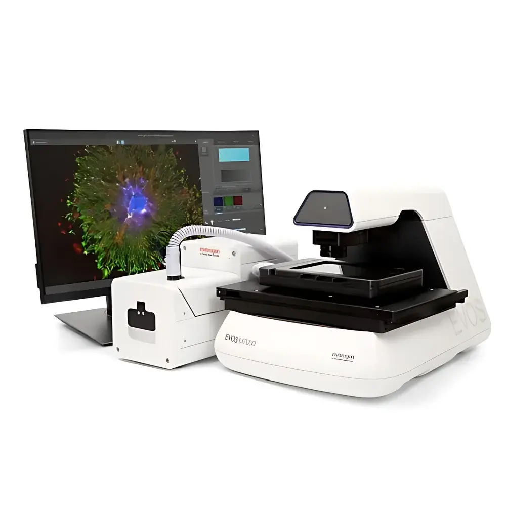
Thermo Fisher Invitrogen EVOS M7000 Automated Live-Cell Imaging System
| Brand | Thermo Fisher |
|---|---|
| Origin | USA |
| Manufacturer Status | Authorized Distributor |
| Origin Category | Imported |
| Model | EVOS M7000 |
| Pricing | Upon Request |
Overview
The Thermo Fisher Invitrogen EVOS M7000 Automated Live-Cell Imaging System is a benchtop, fully integrated platform engineered for high-fidelity, time-resolved optical microscopy of living cells under physiologically relevant conditions. Built upon a modular upright/inverted hybrid optical architecture, the system employs widefield fluorescence and brightfield illumination combined with dual-sensor imaging to simultaneously capture quantitative monochrome fluorescence data and high-fidelity color brightfield morphology. Its core measurement principle relies on precise LED-based excitation, high-numerical-aperture objective optics, and synchronized stage-controlled Z-stack acquisition—enabling non-invasive, longitudinal monitoring of dynamic cellular processes including proliferation, migration, apoptosis, and organelle trafficking. Designed specifically for routine use in core facilities and academic or pharmaceutical research labs, the EVOS M7000 eliminates the need for complex alignment procedures or external environmental chambers by integrating on-stage incubation control directly into the imaging workflow.
Key Features
- High-speed multi-channel imaging: Full 96-well plate scanning across three fluorescence channels completed in under 5 minutes—enabled by rapid LED switching, high-quantum-efficiency monochrome sensor (≥75% QE at 520 nm), and low-latency mechanical stage positioning.
- Modular optical configuration: Supports over 20 interchangeable LED light cubes (365–740 nm excitation range), user-swappable objectives (1.25× to 100×, including phase contrast and long-working-distance options), and standardized vessel adapters for multiwell plates (6–384-well), Petri dishes, chambered coverslips, and custom microfluidic devices.
- Integrated environmental control: On-stage incubation module maintains precise regulation of temperature (±0.2 °C from 20–40 °C), relative humidity (≥95% RH), and gas composition (O2, CO2, N2)—certified for sustained time-lapse imaging up to 72+ hours without medium evaporation or thermal drift.
- Dual-camera imaging architecture: Simultaneous acquisition via a back-illuminated sCMOS monochrome camera (effective pixel size: 6.5 µm; dynamic range: ≥16-bit) for fluorescence quantification and a 5-megapixel RGB CMOS camera (24-bit color depth; CIE-compliant white balance) for contextual brightfield documentation.
- Region-of-interest (ROI) navigation: Seamless transition between low-magnification overview mode (1.25× objective, full-well visualization) and high-resolution tiled scanning (up to 100×) using automated mosaic stitching with sub-pixel registration accuracy.
- Automated acquisition pipeline: Includes hardware-accelerated autofocus (contrast-based + laser-assisted), programmable Z-stack generation (1–100 slices, step sizes 0.1–10 µm), scheduled time-lapse sequences, and batch protocol execution—all controllable via intuitive touchscreen interface or remote API integration.
Sample Compatibility & Compliance
The EVOS M7000 accommodates standard and specialty cell culture formats without modification—including glass-bottom 96-well plates (e.g., MatTek P96G-1.5-5-F), collagen-coated hydrogels, organoid-on-chip devices, and adherent/non-adherent suspension cultures maintained in perfusion-compatible chambers. All optical components comply with ISO 10934-1 (microscope nomenclature) and IEC 61000-6-3 (EMC emissions). Environmental control modules meet ASTM E2927-13 specifications for incubator performance validation. Data acquisition workflows support audit-trail-enabled operation per FDA 21 CFR Part 11 when paired with validated Celleste software deployment and networked domain authentication.
Software & Data Management
System operation and image acquisition are managed through the embedded EVOS Software Suite (v3.5+), featuring real-time preview, multi-dimensional metadata tagging (time, channel, Z-position, environmental parameters), and DICOM-compliant export. Optional Invitrogen Celleste Image Analysis Software (v2.4+) provides FDA-registered tools for object segmentation (adaptive thresholding, machine-learning-assisted nuclei detection), morphometric profiling (cell area, perimeter, circularity, texture entropy), and statistical cohort analysis (ANOVA, linear mixed-effects modeling). Raw TIFF stacks and processed CSV/Excel reports are stored with SHA-256 checksums; local database supports GLP/GMP-aligned project versioning and user-specific access controls.
Applications
- Longitudinal cytotoxicity assessment in drug screening (IC50 kinetics, mitochondrial membrane potential decay)
- Stem cell differentiation tracking via label-free phase contrast + fluorescent reporter co-imaging
- 3D spheroid invasion assays with automated Z-stack reconstruction and volumetric rendering
- CRISPR-edited cell line validation using endogenous fluorescent protein expression dynamics
- Immune synapse formation analysis requiring simultaneous T-cell (CFSE) and target-cell (mCherry) channel registration
- High-content phenotypic profiling for siRNA/shRNA library screens under controlled hypoxia (1–5% O2)
FAQ
Does the EVOS M7000 support oil-immersion objectives?
Yes—select models accommodate 60× and 100× oil-immersion objectives with integrated lens cleaning protocols and refractive index calibration routines.
Can acquisition protocols be exported and shared across multiple instruments?
Yes—EVOS protocols (.evp files) include full hardware configuration, environmental setpoints, and timing logic, and are interoperable across all EVOS M-series platforms running firmware v2.8 or later.
Is Celleste software validated for regulated environments?
Celleste v2.4+ includes a formal validation package compliant with GAMP5 guidelines, supporting IQ/OQ/PQ documentation for use in GLP and GMP laboratories.
What file formats are supported for downstream analysis in third-party tools?
Native export includes OME-TIFF (with complete metadata), ND2 (Nikon), and HDF5 containers; additional plugins enable direct import into Fiji/ImageJ, MATLAB, and Python (via tifffile and ome-zarr libraries).
How is focus stability maintained during extended time-lapse experiments?
The system implements continuous hardware-based focus feedback using a dedicated infrared focus sensor that operates independently of visible-light exposure—ensuring zero phototoxicity-induced focus drift over multi-day acquisitions.


