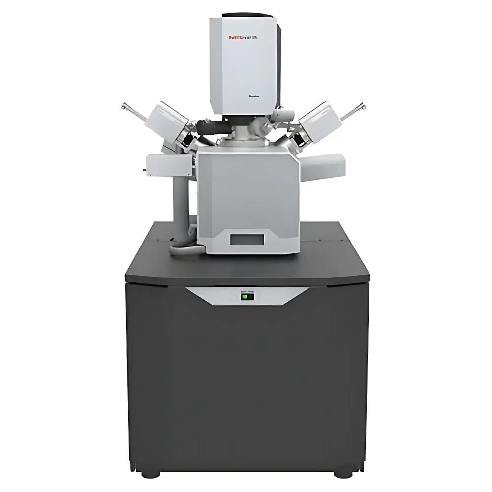
Thermo Fisher Scientific Quattro ESEM Field-Emission Scanning Electron Microscope
| Brand | Thermo Fisher Scientific |
|---|---|
| Origin | Czech Republic |
| Model | Quattro |
| Instrument Type | Floor-standing SEM |
| Electron Source | Cold Field-Emission Gun (CFEG) |
| Magnification Range | 6× to 2,500,000× |
| Accelerating Voltage | 200 V to 30 kV |
| Landing Voltage | 20 eV to 30 keV (with beam deceleration option) |
| Probe Current | 1 pA to 200 nA (continuously adjustable) |
| Secondary Electron Resolution | 1.3 nm @ 30 kV (SE, low-vacuum mode) |
| Backscattered Electron Resolution | 2.5 nm @ 30 kV (BSE, high-vacuum mode) |
| Working Distance for EDS | 10 mm |
| EDS Take-off Angle | 35° |
| Chamber Width | 340 mm |
| Number of Available Ports | 12 (including 3 EDS-compatible ports, two at 180° opposition) |
| Stage Type | 5-axis motorized eucentric stage |
| XY Travel (Quattro C / Quattro S) | 55 × 55 mm / 110 × 110 mm |
| Z Travel | 65 mm |
| Tilt Range | –15° to +90° |
| Rotation | Continuous 360° |
| Maximum Sample Height (from eucentric height) | 85 mm |
| Max Sample Weight | 5 kg (at 0° tilt) |
| Max Sample Diameter (full rotation compatible) | 122 mm |
| Detector Options | In-chamber Everhart-Thornley detector (ETD), Low-Vacuum Detector (LVD), Gas Secondary Electron Detector (GSED), Integrated STEM-in-SEM (WetSTEM™), IR-CCD chamber camera, Nav-Cam™ optical navigation camera |
| Software Platform | 64-bit Windows-based GUI with multi-view display, montage navigation, undo/redo functionality, joystick and multifunction control panel support |
Overview
The Thermo Fisher Scientific Quattro ESEM is a high-performance, floor-standing field-emission scanning electron microscope engineered for maximum analytical flexibility across diverse sample classes—particularly those incompatible with conventional high-vacuum SEM operation. Unlike standard SEMs constrained by stringent vacuum requirements, the Quattro integrates Environmental SEM (ESEM) technology with cold field-emission gun (CFEG) optics, enabling high-resolution imaging and microanalysis under controlled gaseous environments (e.g., water vapor, nitrogen, or CO₂). This capability preserves native hydration states, eliminates charging on insulating specimens, and permits in situ dynamic experiments—including hydration/dehydration, thermal cycling, and contact angle analysis—without conductive coating or cryo-fixation. Its CFEG source delivers exceptional brightness and long-term stability, supporting sub-nanometer resolution in both high-vacuum and low-vacuum modes, while beam deceleration extends landing energy control down to 20 eV—critical for surface-sensitive imaging of beam-sensitive materials.
Key Features
- Triple-vacuum architecture: seamless switching between high vacuum (HV), low vacuum (LV), and environmental vacuum (ESEM) modes—enabling analysis of conductive, insulating, hydrated, thermally unstable, or outgassing samples without preprocessing.
- Cold field-emission electron source with ultra-stable emission current and sub-2 nm resolution at 30 kV (SE, LV/ESEM mode) and 1.0 nm (SE, HV mode).
- Simultaneous multi-signal acquisition: up to four detectors—including ETD, LVD, GSED, and WetSTEM™—can be operated concurrently during a single scan, minimizing electron dose and enabling correlative contrast interpretation (topography, composition, crystallography, cathodoluminescence).
- 5-axis motorized eucentric stage with precision positioning (repeatability <3.0 µm), extended travel (110 × 110 mm XY on Quattro S), ±90° tilt, and continuous 360° rotation—optimized for cross-sectional analysis, grain orientation mapping, and tomographic workflows.
- Modular chamber design with 12 standardized ports (3 dedicated to EDS integration, two positioned at 180° for symmetric geometry), facilitating rapid configuration for EDS, WDS, EBSD, CL, or in situ heating/cooling stages.
- Integrated Nav-Cam™ optical navigation system and IR-CCD chamber camera provide real-time optical context and precise stage-height monitoring—essential for accurate focus, stigmation, and multi-scale correlation.
Sample Compatibility & Compliance
The Quattro accommodates an exceptionally broad sample spectrum—from uncoated polymers, pharmaceutical powders, hydrogels, and plant tissues to geological thin sections, catalytic nanoparticles, and high-temperature superconductors. Its ESEM capability complies with ASTM E1508–22 (Standard Guide for Quantitative Analysis by Energy Dispersive Spectroscopy) and ISO 16700:2016 (Electron probe microanalysis—Quantitative analysis using wavelength dispersive spectrometry), ensuring traceable elemental quantification. The system supports GLP/GMP-aligned workflows through audit-trail-enabled software logging, user-access controls, and full compliance with FDA 21 CFR Part 11 when configured with validated software modules. All vacuum and gas-handling subsystems meet CE and UL safety standards; chamber pressure control adheres to IEC 61000-6-3 for electromagnetic compatibility in laboratory environments.
Software & Data Management
Controlled via a 64-bit Windows-based graphical user interface, the Quattro platform provides a unified environment for instrument control, image acquisition, real-time processing, and data export. The software supports multi-view display (up to four synchronized windows), automated montage stitching, and hierarchical image annotation with metadata tagging (operator, date/time, vacuum mode, detector configuration, beam parameters). Built-in undo/redo functionality ensures experimental reproducibility, while customizable workflow templates accelerate routine analyses—such as particle sizing, phase distribution mapping, or fracture surface assessment. Raw image data are stored in vendor-neutral TIFF format with embedded calibration metadata; spectral datasets (EDS/WDS) comply with the DTSA-II standard for cross-platform compatibility. Optional remote diagnostics and secure cloud backup modules enable centralized fleet management across multi-site laboratories.
Applications
- Materials Science: Characterization of sintered ceramics, metal–matrix composites, and porous battery electrodes under hydrated or heated conditions—without artifact introduction from drying or metallization.
- Life Sciences: Imaging of unfixed, unstained biological tissues, biofilms, and cryo-hydrated cells in near-native state; combined with WetSTEM™ for nanoscale ultrastructure of vitrified thin sections.
- Geosciences: In situ observation of mineral dissolution, clay swelling, and fluid–rock interactions at controlled relative humidity and temperature.
- Pharmaceuticals: Quantitative morphology analysis of inhalable dry powder formulations, tablet coating uniformity, and hydrate phase transitions monitored in real time.
- Failure Analysis: High-fidelity fractography of solder joints, polymer weld lines, and composite delaminations—with simultaneous BSE/SE imaging to distinguish topographic relief from atomic number contrast.
FAQ
What vacuum modes does the Quattro support, and how do they affect imaging performance?
The Quattro operates in three distinct vacuum regimes: high vacuum (≤10⁻⁴ Pa), low vacuum (10–130 Pa), and environmental vacuum (up to 2600 Pa with water vapor or inert gases). Each mode enables specific contrast mechanisms and sample compatibility—high vacuum maximizes resolution for conductive samples; low vacuum mitigates charging on insulators; and ESEM preserves volatile components and enables dynamic environmental experiments.
Can the Quattro perform quantitative EDS analysis in low-vacuum or ESEM mode?
Yes—when equipped with a large-area silicon drift detector (SDD) and optimized geometry (35° take-off angle, 10 mm working distance), quantitative EDS is fully supported in low-vacuum mode per ASTM E1508–22. Performance in ESEM mode depends on gas type and pressure but remains viable for major/minor element mapping with appropriate matrix corrections.
Is the Quattro compatible with third-party in situ stages (e.g., heating, cooling, tensile)?
Absolutely—the 12-port chamber includes standardized electrical, pneumatic, and vacuum interfaces; two EDS-compatible ports at 180° allow symmetrical detector placement during mechanical testing. Thermo Fisher provides mechanical and electrical integration guidelines for certified third-party stages.
How does beam deceleration enhance surface sensitivity?
Beam deceleration reduces landing energy while maintaining high gun extraction voltage—improving signal-to-noise ratio for surface topography and reducing penetration depth. At 20 eV landing energy, interaction volume is confined to the top 1–2 nm, ideal for oxide layer or adsorbate characterization.
What level of software validation is available for regulated environments?
The Quattro software suite can be delivered with IQ/OQ documentation packages and 21 CFR Part 11-compliant audit trail, electronic signatures, and role-based access control—validated in accordance with GAMP 5 principles for pharmaceutical and medical device QA/QC laboratories.


