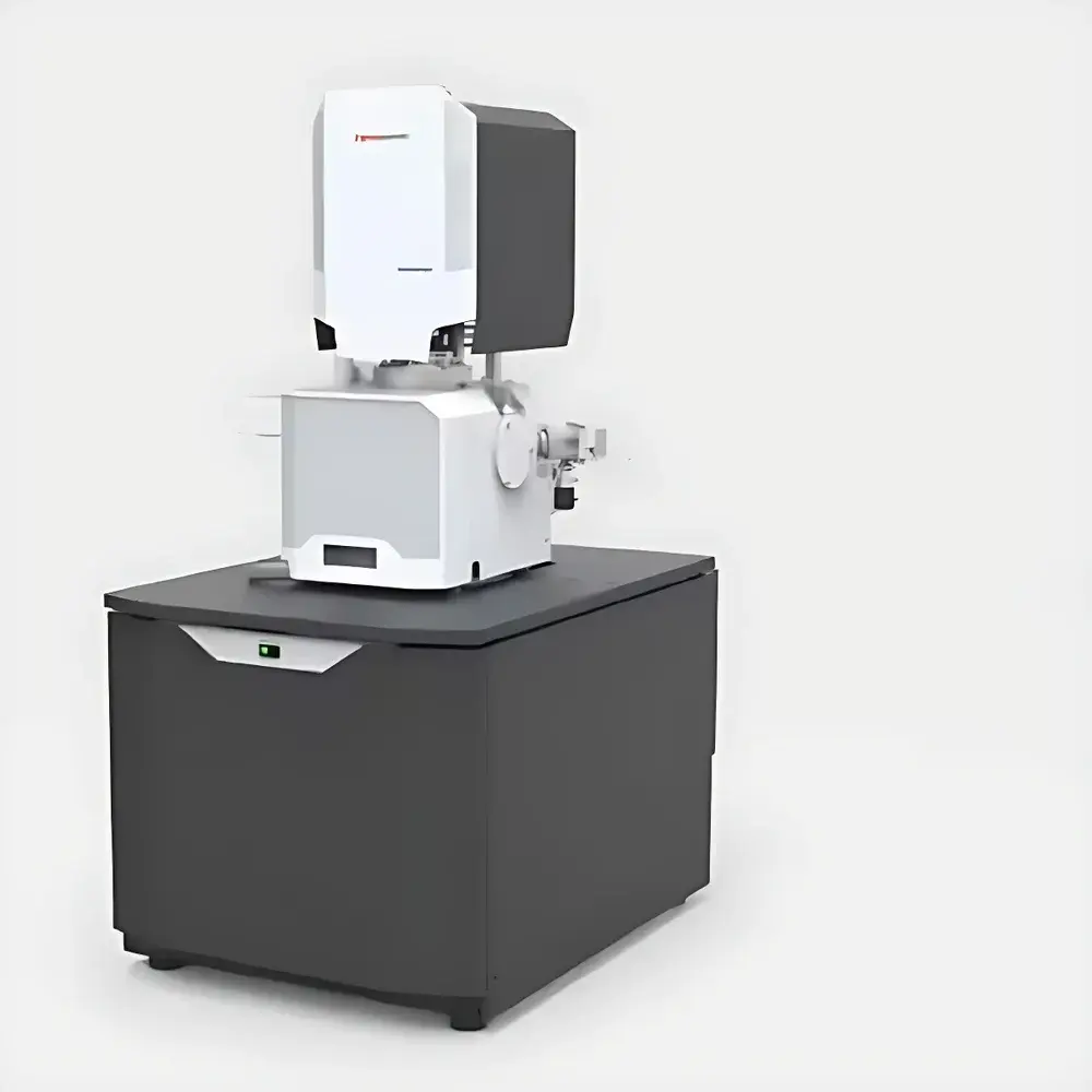
Thermo Fisher VolumeScope 2 SEM Field Emission Scanning Electron Microscope for 3D Serial Block-Face Imaging
| Brand | Thermo Fisher |
|---|---|
| Origin | Shanghai, China |
| Manufacturer Type | Authorized Distributor |
| Origin Category | Domestic (China-manufactured under Thermo Fisher global design & QA) |
| Model | Thermo Fisher VolumeScope 2 SEM |
| Pricing | Available upon Request |
Overview
The Thermo Fisher VolumeScope 2 SEM is a purpose-engineered field emission scanning electron microscope (FE-SEM) optimized for high-fidelity, automated serial block-face (SBF) imaging and isotropic 3D volume reconstruction. Unlike conventional SEMs used for surface topography, the VolumeScope 2 integrates an in-chamber ultramicrotome with a high-stability Schottky field emission gun (FEG), enabling precise in situ sectioning of resin-embedded biological and soft-material specimens under high vacuum — followed immediately by sequential imaging at variable landing energies. This closed-loop SBF-SEM workflow eliminates manual handling, minimizes drift and charging artifacts, and ensures spatial registration across thousands of consecutive sections. Critically, the system implements multi-energy deconvolution — acquiring multiple images per freshly exposed surface at incrementally increased beam voltages (e.g., 500 V to 6 kV), then computationally separating sub-surface signal contributions. This technique overcomes the traditional axial resolution bottleneck imposed by physical section thickness, achieving true isotropic voxel resolution down to 5 × 5 × 10 nm³ in biological tissue and polymer matrices. The result is a quantitative, distortion-corrected 3D dataset suitable for structural phenotyping, connectomics, and multiscale materials characterization.
Key Features
- Integrated in-chamber diamond-knife ultramicrotome with motorized Z-precision stage (sub-10 nm step resolution) and real-time blade wear monitoring.
- High-stability Schottky FEG with ≥24-month guaranteed source lifetime and fully automated gun alignment, beam current stabilization, and aperture heating.
- Dual-mode imaging optics: Immersion-type compound objective lens combining electrostatic and magnetic elements for minimized chromatic aberration and enhanced low-kV performance.
- Beam current range: 1 pA – 400 nA; optimized SBF-SEM operating range: 50 pA – 3.2 nA at landing energies from 500 V to 6 kV.
- Multi-detector configuration: In-lens T1 (low-angle BSE), VS-DBS (variable-pressure BSE), T2 (mid-angle BSE), Everhart-Thornley SE detector (ETD), IR-CCD low-vacuum SE detector, and STEM 3+ segmented annular detector for simultaneous signal channel acquisition.
- Fully oil-free vacuum architecture: Dual turbomolecular pumps (220 L/s + scroll pump + ion getter pumps) achieving <6.3 × 10⁻⁶ mbar chamber pressure within 72 hours; low-vacuum mode supports up to 50 Pa for charge mitigation on non-conductive samples.
Sample Compatibility & Compliance
The VolumeScope 2 accommodates a broad spectrum of embedded and bulk specimens: epoxy- or acrylic-resin-embedded neural tissue (e.g., mouse hippocampus, cortex), cardiac muscle, kidney glomeruli, polymer blends, filtration membranes, battery electrode cross-sections, and composite laminates. Its low-kV imaging capability (down to 20 eV landing energy) and optional low-vacuum detectors enable direct imaging of moderately conductive or beam-sensitive samples without metal coating. All hardware and software modules comply with ISO 9001-certified manufacturing protocols. Data acquisition workflows support audit trails, user authentication, and electronic signatures — aligning with GLP and GMP documentation requirements. While not FDA 21 CFR Part 11–certified out-of-the-box, the system’s Amira Live Tracker and MAPS software platforms are designed for integration into validated laboratory information management systems (LIMS) with configurable metadata logging and version-controlled acquisition scripts.
Software & Data Management
Acquisition and reconstruction are orchestrated via Thermo Scientific MAPS (Microscopy Automation and Processing Suite) and Amira Live Tracker. MAPS provides full automation of SBF cycles — including sectioning depth control, stage repositioning, beam parameter switching, and multi-energy image capture — with scriptable workflows and remote monitoring. Amira Live Tracker enables real-time 2D/3D navigation during acquisition, ROI definition, and adaptive focus correction using continuous beam current feedback. Raw data are stored in standardized HDF5 format with embedded metadata (pixel size, dwell time, kV, detector gain, timestamp). Reconstruction pipelines include deconvolution-based Z-resolution enhancement, nonlinear drift correction, and intensity normalization across thousands of slices. Export formats include TIFF stacks, NRRD, and OME-Zarr for interoperability with Imaris, Fiji/ImageJ, and custom Python-based analysis frameworks.
Applications
- Neuroscience: Isotropic 3D mapping of synaptic vesicles, dendritic spines, and myelinated axons in murine brain tissue at ≤10 nm Z-resolution; compatible with CLEM (correlative light-electron microscopy) workflows.
- Cardiovascular research: Quantitative 3D morphometry of sarcomere organization, mitochondrial cristae density, and intercalated disc ultrastructure in murine myocardium.
- Materials science: Representative volume analysis of phase-separated polymer blends (e.g., PS/PB), pore network topology in asymmetric filtration membranes, and crack propagation pathways in thermoset composites.
- Cell biology: Subcellular organelle segmentation in cryo- or resin-embedded cultured cells; validation of organelle contact sites via volumetric colocalization.
- Quality assurance: Non-destructive 3D defect analysis in microelectronics packaging, solder joint integrity assessment, and additive manufacturing powder bed characterization.
FAQ
What sample preparation methods are recommended for optimal SBF-SEM results?
Standard protocols include chemical fixation (glutaraldehyde/osmium tetroxide), ethanol dehydration, and embedding in Durcupan ACM or Epon resins. For soft polymers, low-viscosity infiltration and slow polymerization minimize shrinkage artifacts.
Can the VolumeScope 2 perform correlative imaging with fluorescence microscopy?
Yes — the system supports fiducial-based registration with light microscopy data when using fluorescent dyes compatible with resin embedding (e.g., Alexa Fluor conjugates stable under osmium treatment).
Is remote operation supported for unattended overnight acquisitions?
Fully supported via secure VPN connection to MAPS; includes automatic error recovery, email/SMS alerts on vacuum loss or knife collision, and real-time progress dashboards.
How is beam damage mitigated during long-duration SBF runs?
Through dynamic beam blanking between sections, dose fractionation strategies, and adaptive dwell time adjustment based on real-time signal-to-noise estimation.
What computing resources are required for deconvolution and reconstruction?
A workstation with ≥64 GB RAM, NVIDIA RTX A6000 GPU, and ≥20 TB of high-throughput storage (NVMe RAID) is recommended for datasets exceeding 1000 slices at 4k × 4k resolution.


