Thermo Scientific™ HeliScan™ Micro-CT System
| Brand | Thermo Fisher |
|---|---|
| Origin | USA |
| Manufacturer Type | Authorized Distributor |
| Origin Category | Imported |
| Model | Thermo Scientific™ HeliScan™ |
| Instrument Form Factor | Floor-standing / Conventional Large-scale |
| Electron Source Type | Cold Field Emission |
| Secondary Electron Image Resolution | Not applicable (X-ray-based micro-CT, not SEM) |
| Magnification Range | Up to 1000× geometric magnification (source-detector geometry dependent) |
| Accelerating Voltage | Not applicable (X-ray tube voltage: 30–160 kV adjustable) |
| Backscattered Electron Image Resolution | Not applicable (X-ray detection system, no electron imaging) |
Overview
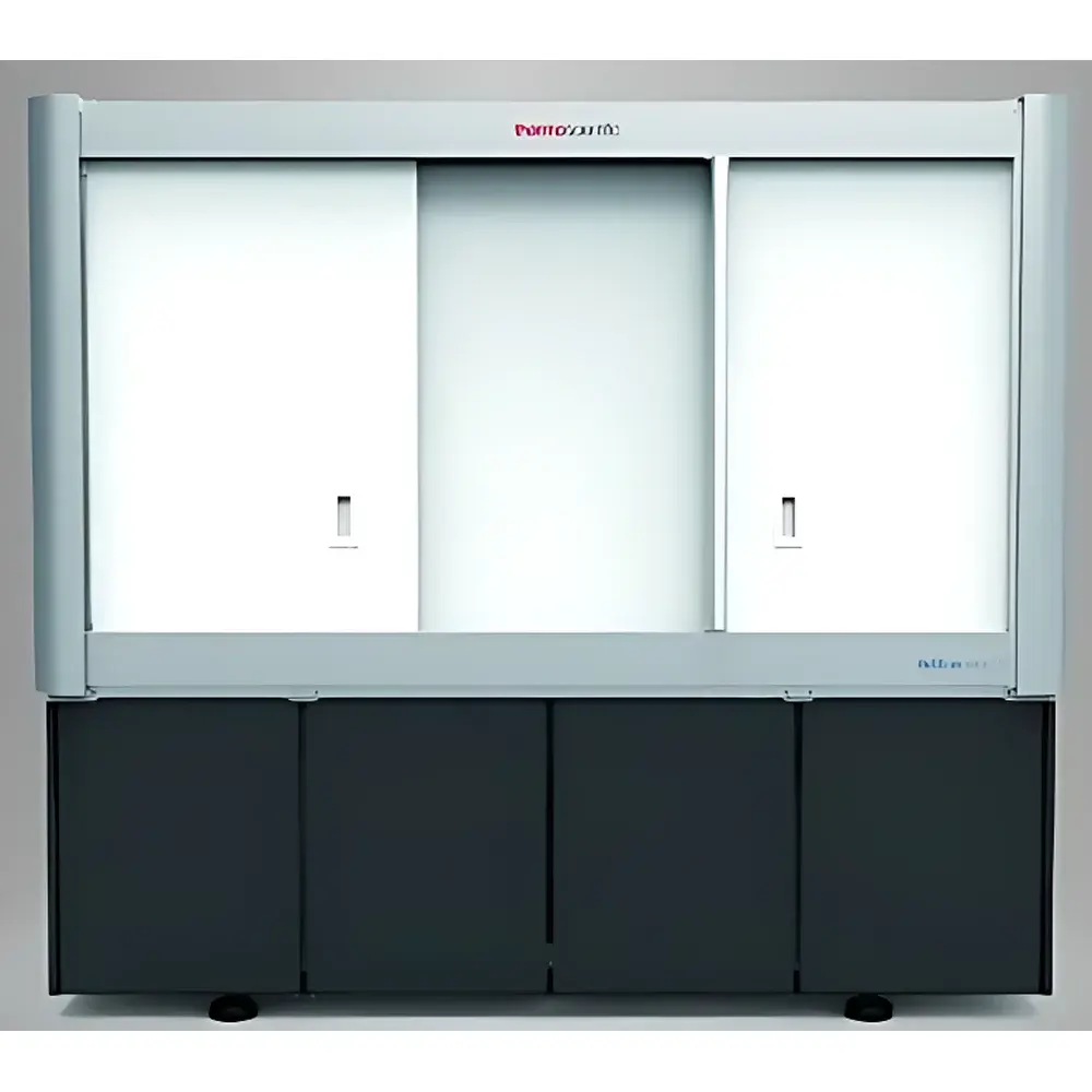
The Thermo Scientific™ HeliScan™ Micro-CT System is a high-performance, floor-standing X-ray computed tomography platform engineered for non-destructive 3D internal structure visualization at micron-scale resolution. Unlike scanning electron microscopes (SEM) or transmission electron microscopes (TEM), the HeliScan operates on the principle of cone-beam X-ray tomography—utilizing a microfocus X-ray source and high-dynamic-range flat-panel detector to acquire projection data across a continuous helical trajectory. This helical acquisition mode, combined with iterative reconstruction algorithms (e.g., SART, OS-SART), enables superior signal-to-noise ratio (SNR), reduced ring and motion artifacts, and enhanced volumetric fidelity compared to conventional step-and-shoot circular scans. Designed for materials science, geoscience, life sciences, and advanced manufacturing, the HeliScan bridges the resolution gap between benchtop micro-CT systems and synchrotron-based imaging—delivering isotropic voxel resolutions down to <1 µm without requiring cryo-fixation or conductive coating.
Key Features
- Helical Trajectory Acquisition: Eliminates stitching artifacts inherent in multi-position circular scans by acquiring projection data in a single, continuous rotation-translation motion—ensuring seamless volumetric reconstruction and improved reproducibility across repeated measurements.
- Auto-Envelope Workflow: Proprietary real-time contour detection algorithm identifies sample boundaries during initial scout scan, automatically optimizing field-of-view, source-to-object distance, and detector exposure—reducing user-dependent setup variability and accelerating workflow throughput.
- Advanced Artifact Suppression: Integrated helix flattening correction compensates for mechanical drift and orbital deviation, mitigating spiral-induced blurring and preserving edge sharpness in reconstructed volumes—critical for quantitative morphometry and phase segmentation.
- Multi-kV X-ray Source: Adjustable tube voltage (30–160 kV) and power (up to 10 W) enable optimal contrast tuning across diverse material densities—from low-Z polymers and biological tissues to high-Z metals and mineral aggregates.
- Modular Detector Architecture: High-resolution, low-noise flat-panel detector with pixel pitch ≤ 50 µm supports both high-speed screening and high-fidelity acquisition modes—scalable to time-resolved (4D) experiments under controlled environmental conditions.
Sample Compatibility & Compliance
The HeliScan accommodates samples up to 150 mm in diameter and 200 mm in height, supporting a broad range of specimen types including rock cores (intact or plug), battery electrodes, additive-manufactured metal parts, composite laminates, and hydrated soft tissues (with appropriate containment). All acquisition protocols comply with ISO 12797 (non-destructive testing — industrial computed tomography), ASTM E1441 (standard guide for computed tomography), and support GLP/GMP-aligned documentation requirements—including audit-trail-enabled acquisition logs, metadata embedding (DICOM-CT compliant), and traceable calibration certificates for geometric and intensity calibration. The system architecture meets FDA 21 CFR Part 11 readiness for electronic records and signatures when deployed with validated Thermo Scientific software configurations.
Software & Data Management
Controlled via Thermo Scientific™ Avizo™ Software (included), the HeliScan provides an integrated pipeline from acquisition to quantification: real-time preview, helical reconstruction, volume rendering, segmentation (threshold-, region-growing-, and AI-assisted), and morphometric analysis (porosity, tortuosity, grain size distribution, connectivity). Raw projections are stored in industry-standard HDF5 format; reconstructed volumes export as DICOM, TIFF stack, or NRRD—ensuring interoperability with third-party platforms such as Dragonfly, MATLAB, or Python-based SciPy/ITK workflows. Data provenance is preserved through embedded metadata (acquisition parameters, timestamps, instrument ID, operator ID), enabling full traceability in regulated environments.
Applications
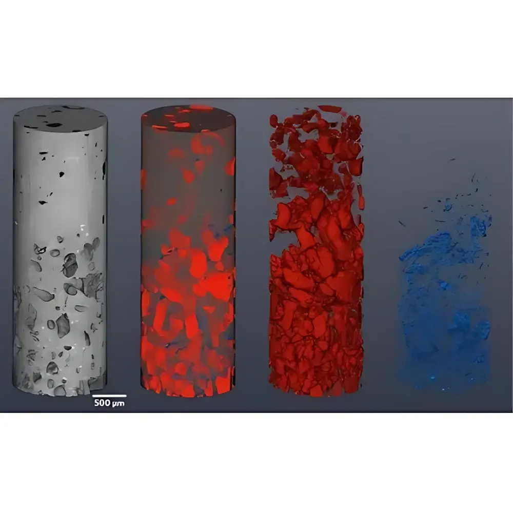
- Petroleum & Geosciences: Quantitative pore-network modeling of reservoir rocks; digital core analysis for permeability prediction; in situ deformation monitoring under triaxial stress; fluid saturation tracking in dynamic drainage/imbibition experiments.
- Materials Science: Defect characterization in castings and welds; porosity mapping in thermal barrier coatings; fiber orientation analysis in carbon-fiber composites; sintering evolution in powder metallurgy.
- Battery Research: Electrode microstructure evolution during cycling; lithium plating detection; separator integrity assessment; solid-electrolyte interphase (SEI) thickness quantification.
- Life Sciences: Bone microarchitecture analysis (trabecular thickness, spacing, connectivity); vascular network reconstruction in ex vivo tissue; developmental morphology in small-animal specimens (e.g., murine lung, zebrafish larvae).
- Additive Manufacturing: As-built defect inventory (lack-of-fusion, keyholing, spatter); dimensional accuracy verification against CAD; residual stress proxy mapping via local density gradients.
FAQ
Is the HeliScan™ a scanning electron microscope (SEM)?
No. The HeliScan is an X-ray micro-computed tomography (micro-CT) system. It does not use electron beams or electron detectors. Its imaging modality is based entirely on X-ray attenuation contrast—not secondary or backscattered electron emission.
What is the typical achievable spatial resolution?
Isotropic voxel sizes range from ~0.5 µm to 10 µm depending on sample size, X-ray energy, detector configuration, and reconstruction parameters. Resolution is governed by geometric magnification and detector pixel size—not by electron optics.
Can the system perform time-resolved (4D) scans?
Yes—when equipped with high-frame-rate detectors and synchronized environmental stages (e.g., heating, compression, fluid injection), the HeliScan supports dynamic in situ and operando studies with temporal resolution down to sub-second intervals per volume.
Does it require vacuum operation?
No. The system operates at ambient air or configurable gas atmospheres (e.g., He, N₂). Vacuum is neither required nor supported, distinguishing it from electron microscopy platforms.
How is calibration maintained for quantitative accuracy?
Geometric calibration is performed using certified phantoms (e.g., tungsten wire grids, glass beads); intensity calibration uses NIST-traceable step wedges. Both are executed during factory commissioning and recommended annually or after major mechanical service.