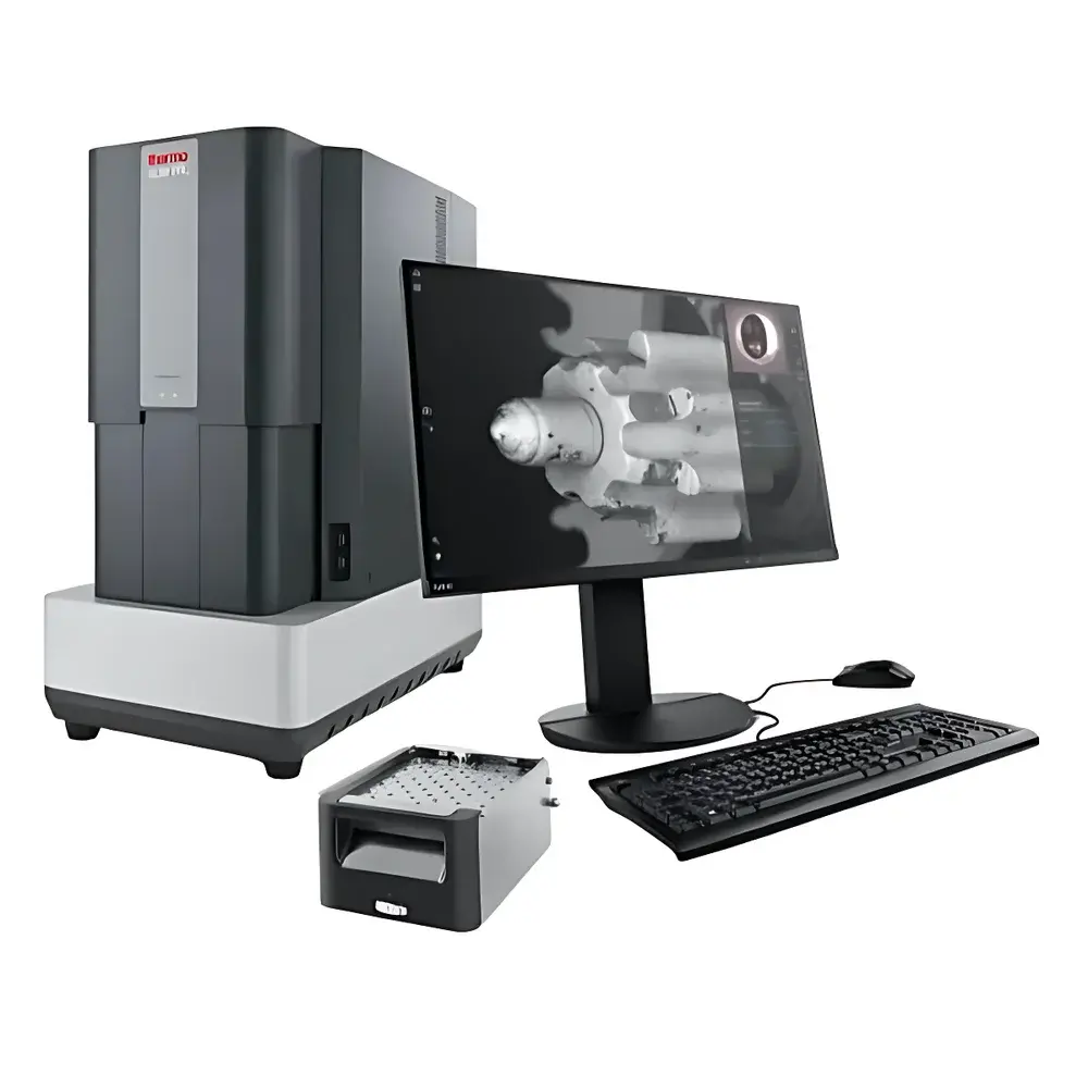
Thermo Scientific Phenom XL G3 Desktop Large-Chamber Automated Scanning Electron Microscope
| Brand | Thermo Scientific Phenom |
|---|---|
| Origin | Netherlands |
| Model | Phenom XL G3 |
| Instrument Type | Desktop SEM |
| Electron Source | CeB6 (Cerium Hexaboride) |
| Secondary Electron Resolution | <8 nm |
| Maximum Magnification | 200,000× |
| Accelerating Voltage Range | 4.8–20.5 kV |
| Backscattered Electron Resolution | <8 nm |
| Optional Detectors | EDS, Secondary Electron Detector |
Overview
The Thermo Scientific Phenom XL G3 is a high-performance desktop scanning electron microscope engineered for industrial quality control, materials R&D, and failure analysis in non-traditional lab environments. Leveraging over 80 years of electron optics heritage—from Philips’ foundational EM100 (1949) through FEI’s pioneering desktop SEM platform (2006) to Thermo Fisher’s current-generation engineering—the XL G3 integrates robust vacuum architecture, a long-life CeB6 thermionic emitter, and intelligent automation into a compact, vibration-tolerant form factor. Unlike conventional SEMs requiring dedicated shielded rooms or floor-mounted foundations, the XL G3 operates reliably in production floors, upper-floor QC labs, mobile laboratories, or field-deployed settings—enabled by its patented internal active damping system and rapid 30-second vacuum cycle. Its core imaging principle relies on raster-scanned electron beam interaction with solid specimens, generating secondary electrons (SE), backscattered electrons (BSE), and characteristic X-rays for simultaneous morphological and compositional analysis—all within a single integrated platform.
Key Features
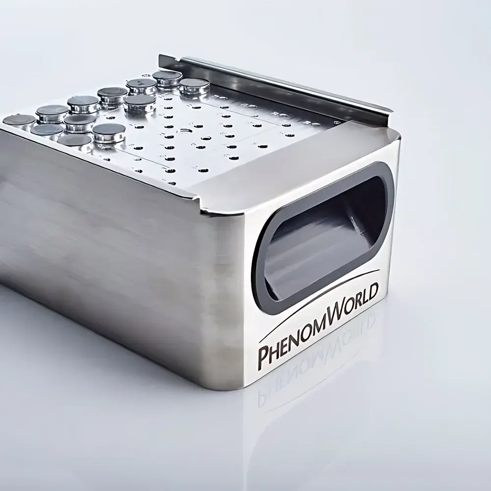
- Desktop-scale large-chamber design accommodating samples up to 100 mm × 100 mm × 50 mm (W × D × H), eliminating the need for time-consuming sectioning or mounting.
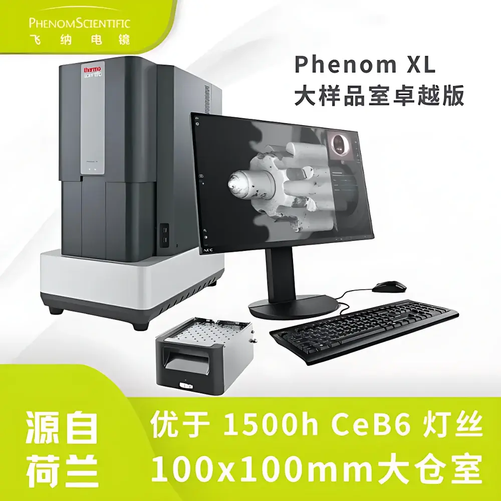
- CeB6 electron source delivering stable, high-brightness emission with >3000 h operational lifetime—reducing filament replacement frequency to approximately once every five years under typical 8 h/day usage.
- Optimized low-voltage imaging capability (down to 2 kV) with enhanced BSE detector sensitivity and improved signal-to-noise ratio, enabling high-fidelity imaging of non-conductive, beam-sensitive, or uncoated specimens—including polymers, biological tissues, ceramics, and solar cell textures—without sputter coating.
- Automated workflow engine supporting fully unattended operation: programmable stage navigation, multi-point acquisition, map-based stitching (Phenom MAPS), and AI-assisted image classification via Python-based Programmable Platform Interface (PPI).
- Integrated EDS-compatible hardware architecture with real-time ChemiSEM color mapping, enabling concurrent topographical and elemental visualization without software switching or session reconfiguration.
Sample Compatibility & Compliance
The Phenom XL G3 supports direct analysis of diverse sample types—including as-received powders, filters, wafers, fracture surfaces, fibers, and coated substrates—without mandatory conductive coating. Its variable-pressure vacuum modes (low/medium/high) accommodate slightly outgassing or hydrated specimens, while the 2–20.5 kV accelerating voltage range ensures optimal contrast and penetration depth across insulating, semi-conductive, and metallic materials. The system complies with ISO/IEC 17025 requirements for testing laboratories when used with documented SOPs; its audit-ready data logging, user-access controls, and timestamped metadata support GLP and GMP workflows. Optional EDS configuration meets ASTM E1508 and ISO 16575 standards for quantitative microanalysis when calibrated per manufacturer protocols.
Software & Data Management
Phenom’s unified software suite eliminates context switching between imaging and analytical modules. ChemiSEM provides live color-coded elemental overlays synchronized with BSE/SE images. ChemiPhase enables phase identification and area-fraction quantification using statistical clustering of EDS spectra. ParticleMetric and FiberMetric automate particle size distribution, shape factor, and fiber length analysis—generating ISO-compliant reports with traceable measurement uncertainty. All acquisitions are stored in a hierarchical digital archive (MAPS), preserving full-resolution image stacks, spectral maps, and stage coordinates. Data export supports TIFF, CSV, HDF5, and industry-standard .emsa formats. Software logs include operator ID, timestamp, instrument parameters, and calibration history—fully compliant with FDA 21 CFR Part 11 when deployed with validated user access management.
Applications
The XL G3 serves critical roles across manufacturing and research domains: semiconductor wafer defect review with automated hotspot localization; battery electrode morphology and SEI layer characterization at low kV; ceramic grain boundary and porosity analysis; polymer fiber diameter uniformity assessment; forensic fiber comparison; environmental filter particulate counting; and metallurgical fracture surface evaluation. Its rapid 1-minute sample-to-image turnaround accelerates QC decision cycles—demonstrated to increase throughput 5–10× versus conventional benchtop SEMs. In R&D, the ability to correlate structure (SE/BSE), composition (EDS), and phase (ChemiPhase) within a single workflow reduces inter-instrument variability and shortens hypothesis-to-validation timelines.
FAQ
Does the Phenom XL G3 require external vibration isolation or dedicated lab space?
No. Its built-in active damping system allows stable operation on standard workbenches—even in high-traffic production areas or upper-floor laboratories.
Can uncoated insulating samples be imaged without charging artifacts?
Yes. Optimized low-kV imaging (2–5 kV), combined with high-efficiency BSE detection and charge compensation algorithms, minimizes charging on polymers, glass, and biological specimens.
Is EDS integration hardware-locked or optional?
EDS is an optional detector module that mounts directly onto the chamber port. It shares the same vacuum and control interface as the base SEM, ensuring seamless co-registration of spectral and morphological data.
What level of automation does the XL G3 support for high-throughput screening?
It supports fully scripted batch analysis of up to 36 samples per load using motorized stage presets, conditional triggers, and Python-based PPI scripting—enabling overnight无人值守 operation.
How is long-term calibration stability maintained?
The CeB6 source provides intrinsic brightness stability; daily auto-alignment routines and drift-compensated stage positioning ensure sub-pixel registration consistency across multi-day mapping sessions.


