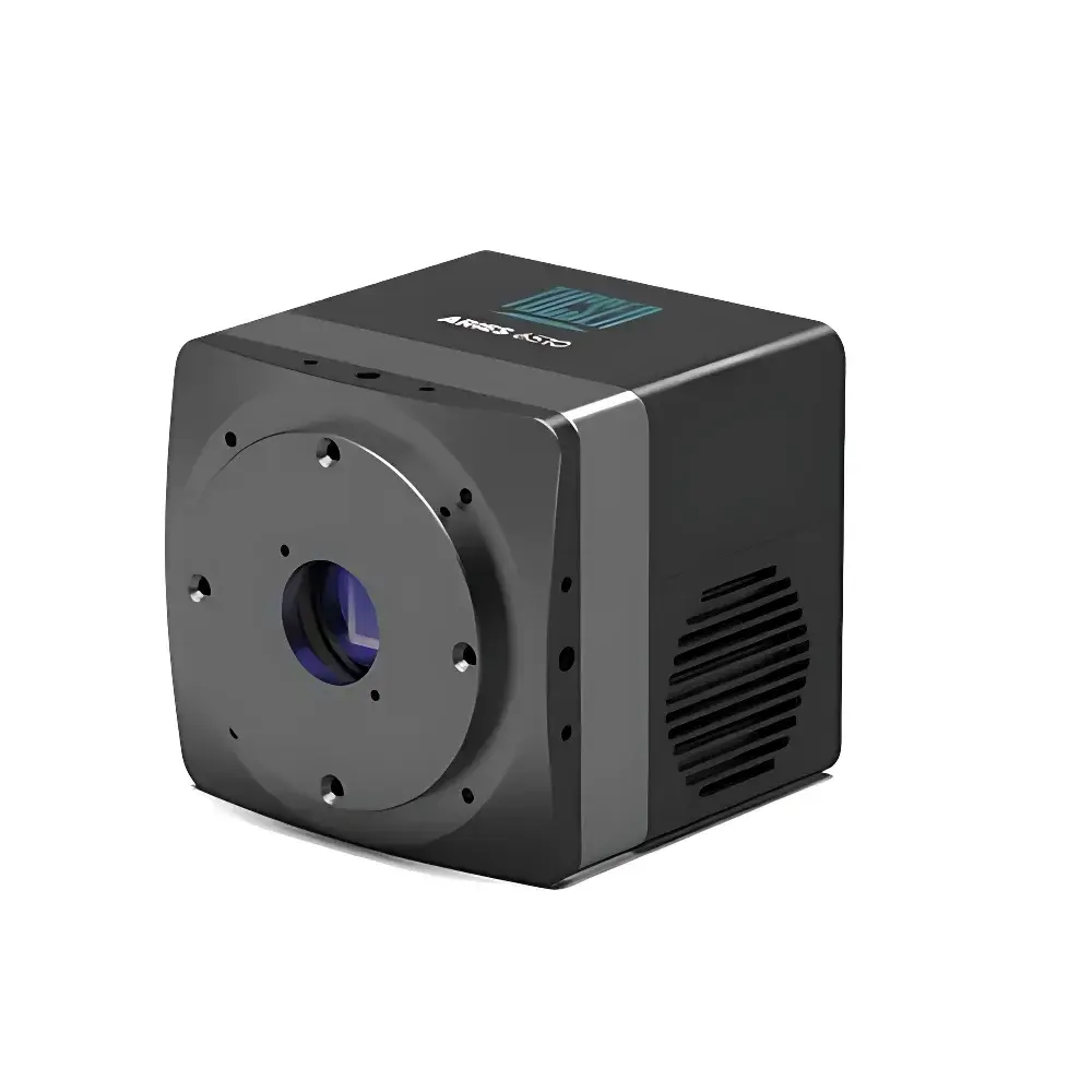
TUCSEN Aries 6506 BSI sCMOS Microscopy Camera
| Brand | TUCSEN |
|---|---|
| Origin | Fujian, China |
| Manufacturer Type | OEM Manufacturer |
| Product Category | Domestic |
| Model | Aries 6506 |
| Image Resolution | 2400 (H) × 2400 (V) |
| Pixel Size | 6.5 µm × 6.5 µm |
| Sensor Active Area | 15.7 mm × 15.7 mm |
| Diagonal Field of View | 22 mm |
| Readout Speeds | 6.9 fps / 111 fps / 117 fps / 200 fps |
| Quantum Efficiency | 95% (peak) |
| Read Noise | 0.7 e⁻ (Super-Sensitivity mode) |
| Full Well Capacity | 1,000 e⁻ or 15,000 e⁻ selectable per readout mode |
| Bit Depth | 11–16 bit (mode-dependent) |
| Cooling | Air-cooled to 0°C (ambient 25°C) / Water-cooled to −10°C (coolant 20°C) |
| Interface | Dual 10GigE |
| Mount | C-Mount |
| Power | 12 V / 8 A |
| Max Power Consumption | ≤55 W |
| Dimensions | 95 mm (H) × 100 mm (W) × 100 mm (L) |
Overview
The TUCSEN Aries 6506 is a high-performance back-illuminated scientific CMOS (BSI sCMOS) camera engineered for demanding quantitative microscopy applications—including live-cell calcium imaging, time-lapse morphological tracking, high-throughput slide scanning, and digital pathology workflows. Built around the Gpixel GSENSE6510BSI sensor, it delivers an industry-leading peak quantum efficiency of 95% and ultra-low read noise down to 0.7 e⁻ in Super-Sensitivity mode—enabling photon-limited detection without compromising temporal resolution. Its 2400 × 2400 pixel array, with 6.5 µm pixels and a 22 mm diagonal field of view, is optimized for standard C-mount microscope integration while maintaining compatibility with wide-field, confocal, and light-sheet platforms. The camera operates on a dual 10 Gigabit Ethernet interface, eliminating dependency on proprietary frame grabbers and supporting deterministic, low-latency data streaming in multi-camera or synchronized acquisition environments.
Key Features
- Back-Illuminated sCMOS Architecture: Maximizes photon collection efficiency across visible to near-UV wavelengths (350–1100 nm), critical for low-light fluorescence, FRET, and Ca²⁺ indicator dyes (e.g., GCaMP, Fluo-4).
- Four Configurable Readout Modes: HDR (high dynamic range), Speed (200 fps at reduced FOV), Sensitivity (117 fps, balanced SNR), and Super-Sensitivity (6.9 fps, 0.7 e⁻ noise, 15,000 e⁻ full-well)—each with independent bit-depth (11–16 bit) and gain calibration.
- Thermal Management Flexibility: Dual cooling options—air-cooling to 0°C (ΔT = −25°C vs. 25°C ambient) or water-cooling to −10°C—ensure stable dark current (<0.5 e⁻/pix/s at 0°C), essential for long-exposure Z-stacks and quantitative intensity calibration.
- GigE Vision 2.0 Compliance: Fully compliant with EMVA1288 standards; supports GenICam XML configuration, hardware-triggered exposure synchronization (global reset & rolling shutter), and precise timestamping via PTPv2 (IEEE 1588) for multi-modal system alignment.
- FPGA-Accelerated Onboard Processing: Real-time defect pixel correction (DPC), region-of-interest (ROI) cropping, binning (2×2, 4×4), and histogram-based auto-exposure—all executed without host CPU overhead.
Sample Compatibility & Compliance
The Aries 6506 supports diverse biological sample formats—from single adherent cells in chambered coverslips to whole-tissue sections on glass slides (up to 15 × 15 cm with motorized stage integration). Its 16-bit linear response and calibrated gain/offset tables enable absolute intensity quantification traceable to NIST-traceable photodiode references. For regulated environments, the camera’s deterministic trigger latency (<1 µs jitter), audit-log-capable SDK, and metadata-embedded TIFF/ND2 export support compliance with GLP/GMP documentation requirements. While not FDA-cleared as a medical device, its performance parameters align with ISO 10940 (microscope camera characterization) and ASTM E2811 (digital pathology scanner validation guidelines) for research-use-only (RUO) deployment.
Software & Data Management
TUCSEN provides a cross-platform SDK (C/C++, C#, Python bindings) with full GenICam compliance, enabling seamless integration into custom acquisition pipelines (e.g., Micro-Manager, MATLAB Image Acquisition Toolbox, or LabVIEW). Native support for HDF5 and OME-TIFF ensures metadata-rich, multi-dimensional storage compatible with Bio-Formats and QuPath. Time-series acquisitions include embedded frame timestamps, temperature logs, and exposure parameter history—facilitating reproducible analysis under FAIR (Findable, Accessible, Interoperable, Reusable) data principles. Optional TUCSEN Studio software offers real-time spectral unmixing, drift correction, and batch-processed focus stacking for pathology slide digitization workflows.
Applications
- Live-Cell Calcium Imaging: Sub-second temporal resolution combined with sub-electron read noise enables reliable detection of transient Ca²⁺ spikes in neuronal cultures or cardiomyocytes under physiological illumination intensities.
- High-Throughput Screening: 200 fps burst acquisition at cropped ROI supports kinetic assays (e.g., ion channel pharmacology) across 96-/384-well plates when paired with automated stages.
- Digital Pathology Scanning: 22 mm diagonal FOV reduces tile count by ~40% versus conventional 16 mm sensors—accelerating whole-slide imaging (WSI) without sacrificing resolution or SNR.
- Multiplexed Immunofluorescence: High QE and low crosstalk facilitate simultaneous detection of spectrally overlapping probes (e.g., Cy3/Cy5/Cy7) using narrow-band filter sets and spectral unmixing algorithms.
- Light-Sheet Microscopy: Low-latency triggering and global reset capability ensure precise synchronization with pulsed illumination, minimizing motion blur in cleared-tissue volumetric imaging.
FAQ
Is the Aries 6506 compatible with third-party microscope control software?
Yes—the camera fully supports GenICam-compliant platforms including Micro-Manager 2.0, Thorlabs Kinesis, and Andor Solis via standardized XML feature descriptions and GigE Vision streaming protocols.
What cooling method is recommended for long-duration time-lapse experiments?
Water-cooling to −10°C is advised for acquisitions exceeding 30 minutes to maintain thermal stability and suppress dark current drift below 0.1 e⁻/pix/s.
Does the SDK support FDA 21 CFR Part 11 compliance features?
The SDK includes optional electronic signature logging, user-access controls, and immutable acquisition metadata—configurable to meet Part 11 requirements when deployed within validated laboratory information management systems (LIMS).
Can the camera operate in global shutter mode?
No—it uses a highly optimized rolling shutter architecture with global reset functionality, providing effective synchronization for most fluorescence microscopy modalities while retaining superior QE and noise performance.
How is calibration data handled during multi-camera setups?
Each unit ships with factory-measured EMVA1288 reports (including PRNU, DSNU, and linearity curves); these are loaded automatically by the SDK to normalize intensity across synchronized cameras in array-based light-sheet or multi-angle imaging rigs.


