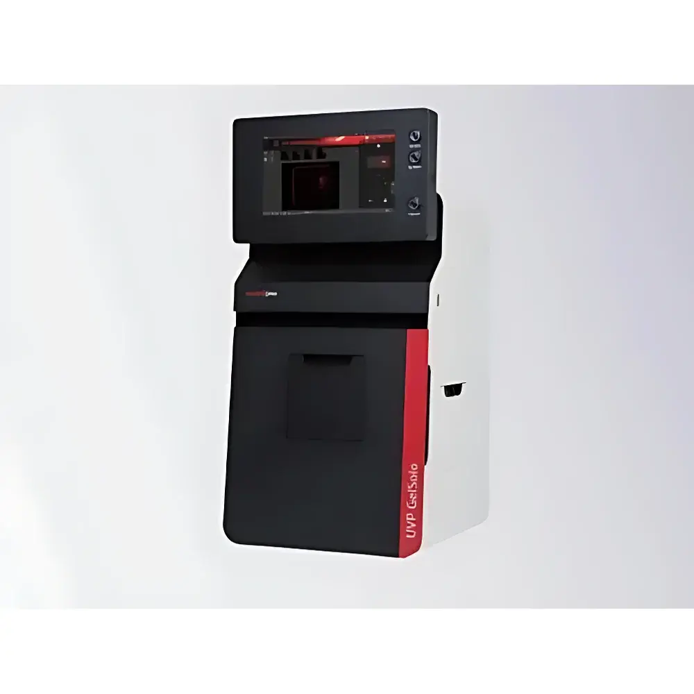
UVP GelSolo Independent Gel Documentation System
| Brand | Analytik Jena |
|---|---|
| Origin | Germany |
| Model | UVP GelSolo |
| Instrument Type | Standalone Gel Imaging System |
| CCD Resolution | 5.0 Megapixels |
| CCD Sensor Size | 2/3" |
| Lens | 8–48 mm, f/1.2 |
| Light Sources | Transmitted UV (302 nm), Transmitted Blue Light (470 nm), Transmitted White Light, Reflected White Light, Reflected Blue Light |
| Standard Filters | Ethidium Bromide (EB) |
| Optional Filters | SYBR® Green, SYBR® Gold, Coomassie Blue, Silver Stain |
| Display | 11.6-inch Capacitive Touchscreen |
| Image Storage | Internal SSD (JPG, TIFF, PNG) |
| Connectivity | Ethernet, Wi-Fi, USB |
| Safety Features | UV-Blocking Observation Window, Four-Door Access Design, Blue-Light Conversion Plate (optional) |
Overview
The UVP GelSolo Independent Gel Documentation System is a fully self-contained, benchtop gel imaging platform engineered for precision, safety, and operational autonomy in molecular biology laboratories. Unlike PC-dependent systems, the GelSolo integrates a high-performance 5.0-megapixel scientific-grade CCD sensor (2/3″ format), a motorized f/1.2 zoom lens (8–48 mm), and an embedded Linux-based imaging engine—enabling real-time acquisition, visualization, annotation, and analysis without external computing hardware. Its core measurement principle relies on quantitative fluorescence and absorbance detection across multiple excitation/emission bands, optimized for nucleic acid (e.g., DNA/RNA gels stained with EB, SYBR® Green, or SYBR® Gold) and protein applications (Coomassie-stained SDS-PAGE, silver-stained gels, DAB-developed Western blots). The system’s optical architecture supports both trans-illumination and epi-illumination modalities, ensuring consistent signal-to-noise ratios across diverse sample types and staining chemistries.
Key Features
- Standalone operation via integrated 11.6-inch capacitive touchscreen interface—no PC required for image capture, preview, or basic quantification
- Five independently controllable light sources: transmitted UV (302 nm), transmitted blue light (470 nm), transmitted white light, reflected white light, and reflected blue light—enabling broad compatibility with legacy and modern dyes
- UV-blocking observation window with >99.9% attenuation of 254–365 nm radiation, compliant with IEC 62471 photobiological safety standards for Class 1 LED/UV equipment
- Four-door access design with interlocked UV shutters—meets ISO 15223-1 labeling requirements for medical device safety and facilitates rapid, contamination-controlled gel excision
- Optional blue-light conversion plate eliminates direct UV exposure during gel documentation; compatible with blue-light–optimized dyes (e.g., SYBR® Safe), reducing photodamage to DNA and eliminating ethidium bromide handling per CLSI MM18-A guidelines
- f/1.2 variable-aperture lens ensures high photon collection efficiency—even under low-fluorescence conditions—while maintaining uniform field illumination and minimal geometric distortion
Sample Compatibility & Compliance
The GelSolo supports standard electrophoretic formats including agarose (0.5–3.0%), polyacrylamide (8–20%), and gradient gels. It accommodates mini- and midi-format gels (up to 15 × 15 cm), blot membranes (PVDF, nitrocellulose), and tissue sections. All optical filters comply with ISO 11146-2 spectral bandwidth tolerances and are certified for peak transmission >85% at designated emission maxima (e.g., 590 ± 10 nm for EB, 520 ± 10 nm for SYBR® Green). The system adheres to EU Directive 2014/30/EU (EMC), 2014/35/EU (LVD), and carries CE marking for in vitro diagnostic use under IVDR Annex XVI. For GLP/GMP environments, audit trails, user access controls, and electronic signature support are available via optional software modules aligned with FDA 21 CFR Part 11 requirements.
Software & Data Management
The embedded GelSolo OS includes multilingual UI (English, German, French, Spanish, Chinese) and native support for TIFF, JPEG, PNG, and RAW (16-bit) export. Image analysis functions include lane/profile tracing, molecular weight estimation (with reference ladder calibration), band intensity quantification (background subtraction, volume integration), and % saturation reporting. Data export supports DICOM-SR metadata embedding for LIMS integration. Images are stored on internal encrypted SSD and accessible via SMB/CIFS over Ethernet or IEEE 802.11ac Wi-Fi. Optional USB 3.0 host port enables direct connection to thermal printers (e.g., Brother QL-1110NWB) for GLP-compliant hardcopy generation with timestamp, operator ID, and instrument serial number watermarking.
Applications
- Quantitative analysis of PCR products, restriction digests, and plasmid preparations on agarose gels
- Detection and densitometry of Coomassie- and silver-stained protein gels for expression profiling
- Documentation of chemiluminescent and chromogenic Western blots (DAB, BCIP/NBT)
- Validation of CRISPR editing efficiency via T7E1 or Surveyor nuclease assays
- Preparative gel excision under blue-light illumination for downstream cloning or mass spectrometry
- Teaching laboratory use—intuitive interface reduces training time while maintaining traceable data integrity
FAQ
Does the GelSolo require a computer for routine imaging tasks?
No—the system operates entirely independently using its onboard processor and touchscreen interface. A PC is only needed for advanced batch analysis or LIMS integration.
Can the GelSolo be validated for regulated environments (e.g., clinical labs)?
Yes—when configured with optional 21 CFR Part 11-compliant software, it supports electronic signatures, audit trails, and role-based access control required for CLIA- and CAP-accredited facilities.
What safety certifications does the UV illumination module hold?
The UV source meets IEC 62471 (Photobiological Safety of Lamps) Class 1 requirements and incorporates dual redundant shutter interlocks per EN 60825-1 for laser product safety equivalence.
Is SYBR® Gold compatible with the standard filter set?
The default EB filter is not optimal for SYBR® Gold. An optional SYBR® Gold-specific emission filter (peak transmission at 530 ± 10 nm) is available and factory-calibrated for quantitative linearity across 0.1–100 ng DNA ranges.
How is image data backed up and archived?
Images are automatically saved to internal storage with SHA-256 checksums. Scheduled backups can be configured to network-attached storage (NAS) via SMB or to cloud endpoints (AWS S3, Azure Blob) using TLS 1.2–encrypted REST API calls.
Related Products