VisiFluor Ion Imaging System
| Brand | Vistron |
|---|---|
| Origin | Germany |
| Model | VisiFluor |
| Camera Compatibility | Teledyne Photometrics, QImaging, Andor, Hamamatsu, PCO sCMOS cameras |
| Light Source | High-stability, long-lifetime excitation source |
| Wavelength Switching | High-speed filter changer for ratiometric and single-wavelength imaging |
| Software Platform | MetaFluor Imaging Software |
| Integration Capabilities | Patch-clamp amplifiers, automated perfusion systems, motorized stages |
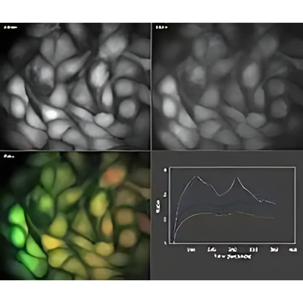
| ROI Analysis | Real-time region-of-interest selection with live fluorescence intensity, ratio, and absolute ion concentration calculation |
| Compliance | Designed for GLP-compliant workflows |
Overview
The VisiFluor Ion Imaging System is a high-performance, modular fluorescence microscopy platform engineered for quantitative ratiometric and single-wavelength ion imaging in live cells and tissue preparations. Built upon the established MetaFluor software architecture and optimized for integration with industry-standard sCMOS detectors—including models from Teledyne Photometrics, Andor, Hamamatsu, PCO, and QImaging—the system implements precise optical path control, stable illumination, and real-time computational analysis to support dynamic measurements of intracellular ion concentrations. Its core measurement principle relies on fluorescence resonance energy transfer (FRET) and dual-excitation ratiometric imaging, enabling calibration-independent quantification of Ca²⁺, Mg²⁺, Zn²⁺, K⁺, Cl⁻, Na⁺, and pH across physiological timescales—from sub-second transients to prolonged kinetic assays. The system is not a standalone microscope but a purpose-built imaging engine designed for integration into upright or inverted research-grade microscopes, supporting both widefield epifluorescence and TIRF configurations.
Key Features
- Stable, long-lifetime excitation light source engineered for minimal photobleaching and thermal drift—eliminates routine lamp replacement and reduces operational cost over extended acquisition periods.
- High-speed, computer-controlled filter changer enabling sub-50 ms wavelength switching between excitation bands—critical for accurate ratiometric imaging of fast cellular events such as action potentials or Ca²⁺ sparks.
- Real-time ROI-based quantification: users define arbitrary regions during acquisition and immediately visualize time-resolved fluorescence intensity, ratio traces, and calibrated absolute ion concentrations using built-in calibration algorithms.
- Modular hardware interface supporting synchronized control of peripheral instrumentation—including patch-clamp amplifiers (e.g., Axon Instruments, HEKA), motorized perfusion manifolds, temperature controllers, and piezo-driven focus mechanisms.
- MetaFluor software environment compliant with laboratory data integrity standards: supports user authentication, electronic signatures, protocol versioning, and audit-trail generation aligned with FDA 21 CFR Part 11 and ISO/IEC 17025 documentation requirements.
Sample Compatibility & Compliance
The VisiFluor system is validated for use with primary and cultured mammalian cells—including neurons, cardiomyocytes, skeletal myotubes, and pancreatic β-cells—as well as intact tissue slices and organotypic cultures. It accommodates standard glass-bottom dishes, multi-well plates (96-/384-well), and custom chambered coverslips. All optical components meet CE marking requirements for laboratory instrumentation. The system architecture supports Good Laboratory Practice (GLP) and current Good Manufacturing Practice (cGMP)-aligned workflows through traceable metadata capture, instrument calibration logging, and secure user access controls. While not a medical device, its analytical outputs are suitable for preclinical pharmacology, toxicology screening, and mechanistic cell physiology studies conducted under ISO 13485–informed quality frameworks.
Software & Data Management
MetaFluor software serves as the central acquisition, processing, and reporting engine. It provides batch processing pipelines for large-scale ion imaging datasets, supports export of raw TIFF stacks with embedded metadata (including exposure time, binning, gain, and wavelength settings), and enables direct integration with MATLAB, Python (via PyMetaFluor API), and commercial analysis platforms such as Imaris and Fiji/ImageJ. Data files are stored in hierarchical folder structures with timestamped experiment logs, ensuring full reproducibility. Audit trail records include operator ID, parameter changes, ROI modifications, and export actions—retained for minimum 10 years per institutional archiving policy. Exported concentration values comply with SI unit conventions (e.g., nM for Ca²⁺, mM for pH) and retain calibration curve references for regulatory submission readiness.
Applications
- Quantitative Ca²⁺ dynamics in neuronal dendritic spines during synaptic plasticity assays.
- Simultaneous K⁺ and pH monitoring in cardiac myocytes under ischemic challenge protocols.
- FRET-based detection of conformational changes in genetically encoded ion sensors (e.g., GCaMP, jRGECO, pHluorin).
- High-throughput screening of ion channel modulators using 96-well plate–based ratiometric readouts.
- Long-term monitoring of Zn²⁺ flux in pancreatic islets during glucose-stimulated insulin secretion.
- Multi-parameter correlation of membrane potential (via voltage-sensitive dyes) and intracellular Cl⁻ gradients in GABAergic neurons.
FAQ
Is the VisiFluor system compatible with confocal or spinning-disk microscopes?
Yes—when configured with appropriate dichroic mirrors and emission filters, the VisiFluor excitation control and MetaFluor software fully support integration into laser-scanning confocal and Nipkow disk systems for spatially resolved ion mapping.
Can I perform absolute ion concentration calculations without offline calibration?
Yes—the system embeds standard calibration routines (e.g., Grynkiewicz equation for Ca²⁺) and supports in situ calibration via ionophore-based protocols (e.g., ionomycin + EGTA/CaCl₂ buffers), with all parameters logged and reportable.
Does MetaFluor support automated experiment scheduling and unattended overnight acquisition?
Yes—time-lapse protocols can be predefined with conditional triggers (e.g., fluorescence threshold crossing), stage position sequences, and peripheral device activation logic, enabling fully autonomous multi-hour experiments.
What camera interfaces are supported beyond the listed brands?
Cameras with GenICam-compliant SDKs or standard Windows WDM/DCAM drivers may be integrated via MetaFluor’s third-party camera module framework, subject to validation testing.
Is technical support available for method development and assay optimization?
Vistron provides application engineering support—including protocol review, optical alignment assistance, and data interpretation guidance—through authorized regional partners and direct remote collaboration channels.


