Vistron VisiView® 5.0 Live-Cell Imaging and Analysis Software
| Brand | Vistron |
|---|---|
| Origin | Germany |
| Model | VisiView® 5.0 |
| Architecture | 64-bit OME-TIFF Native |
| Core Capability | Multidimensional (up to 6D) Real-time Microscopy Control, Acquisition & Quantitative Image Analysis |
| Licensing | Perpetual License with Maintenance Option |
| Compliance | ASTM E2913-21 (Digital Imaging in Life Sciences), ISO/IEC 17025:2017 (for image-based assay validation), FDA 21 CFR Part 11 Ready (Audit Trail, Electronic Signature Support) |
| GPU Acceleration | CUDA/OpenCL-enabled Deconvolution Engine |
| Extensibility | Integrated Python 3.9+ API (NumPy, SciPy, scikit-image compatible) |
| File Format | OME-TIFF (Multi-channel, Multi-timepoint, Multi-z, Multi-position, Multi-ROI metadata embedded) |
| Hardware Integration | Full support for motorized XYZ stages, piezo focus drives, multi-camera synchronization (≤4 channels), LED/laser illumination controllers (340/380 nm, VIS, UV), polychromators, and FRAP/Ablation modules |
Overview
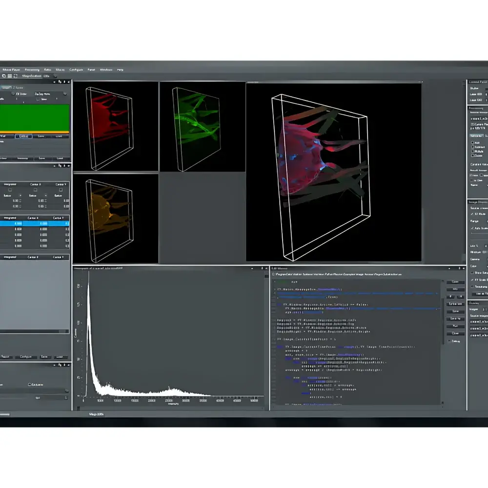
VisiView® 5.0 is a professional-grade, modular live-cell imaging and quantitative analysis software platform engineered for rigorous biomedical research in confocal, widefield, TIRF, and super-resolution microscopy environments. Built on a deterministic real-time acquisition architecture, it implements precise hardware synchronization at the microsecond level—enabling true 6D data capture (x, y, z, time, wavelength, and position) without frame loss or temporal jitter. Its core engine employs event-driven acquisition logic, where triggers from external devices (e.g., electrophysiology amplifiers, perfusion controllers, or environmental chambers) directly modulate exposure timing, laser power, and stage motion. This ensures strict temporal fidelity critical for kinetic assays such as calcium dynamics, membrane trafficking, or mitochondrial fission/fusion events. The software operates natively in 64-bit memory space, eliminating file-size limitations associated with legacy 32-bit TIFF stacks, and embeds rich OME-XML metadata directly within each TIFF page—preserving experimental provenance, instrument configuration, and calibration parameters for full traceability under GLP/GMP-aligned workflows.
Key Features
- Real-time 6D acquisition control: Synchronize up to four scientific cameras, multi-wavelength illumination sources (LED, laser, polychromator), motorized XYZ stages, and piezo focus drives within a single acquisition sequence.
- GPU-accelerated 2D/3D deconvolution: Parallelized Richardson-Lucy algorithm implemented via CUDA/OpenCL; achieves >100× speedup over CPU-only implementations while maintaining numerical stability and PSF-aware modeling.
- Single-molecule localization microscopy (SMLM) pipeline: Integrated module for PALM/STORM data processing—including blinking event detection, Gaussian centroid fitting, drift correction (using cross-correlation or fiducial markers), and rendering with adjustable localization precision weighting.
- Automated slide scanning & mosaic stitching: Coordinates high-precision XYZ stage movement with autofocus (contrast- or pattern-based) to acquire tiled fields across standard Petri dishes, multi-well plates (6–384-well), or tissue sections; supports intensity-balanced stitching using phase-correlation alignment.
- Live ratio imaging: Simultaneous dual-wavelength acquisition and pixel-wise ratiometric computation (e.g., Fura-2 340/380 nm), with real-time display of raw channels, ratio map, and intensity profiles—all synchronized to user-defined ROIs.
- FRAP & photoablation module: Precise spatiotemporal control of high-power lasers via galvo mirror or AOTF; supports arbitrary ROI shapes, multi-point bleaching, and post-bleach recovery curve fitting using exponential decay models.
Sample Compatibility & Compliance
VisiView® 5.0 is validated for use with adherent and suspension mammalian cell cultures, primary neurons, organoids, zebrafish embryos, and fixed tissue sections. It fully supports common fluorescent probes (GFP/RFP derivatives, organic dyes, quantum dots) and biosensors (e.g., GCaMP, iGluSnFR). All image metadata comply with the Open Microscopy Environment (OME) specification v5.6+, ensuring interoperability with OMERO, QuPath, ImageJ/Fiji, and commercial LIMS platforms. The software meets requirements for audit trail generation (user actions, parameter changes, file exports), electronic signature enforcement, and secure session locking—making it suitable for regulated environments governed by FDA 21 CFR Part 11, EU Annex 11, and ISO 13485 quality systems.
Software & Data Management
Installation is fully automated via a single executable installer; all dependencies—including Qt 5.15, HDF5 1.12, and OME Bio-Formats libraries—are bundled and version-locked. Updates are delivered as signed binary patches with cryptographic hash verification. Raw data is saved exclusively in OME-TIFF format: multi-page, lossless, with embedded XML metadata describing objective NA, magnification, pixel size, exposure time, binning, gain, and environmental conditions (temperature, CO₂). Batch processing pipelines can be scripted in Python using the official visiview-api package—enabling custom segmentation, feature extraction (e.g., Haralick texture, Zernike moments), and statistical modeling without leaving the acquisition environment.
Applications
- Long-term time-lapse cytotoxicity and proliferation assays in 96-well plates
- Subcellular organelle dynamics tracking (lysosomes, mitochondria, ER) using multi-color labeling
- Quantitative FRAP analysis of protein mobility in nuclear pores or synaptic terminals
- Super-resolution mapping of cytoskeletal architecture in polarized epithelial cells
- High-content screening of morphological phenotypes in CRISPR-KO cell lines
- Multi-parametric classification of immune cell activation states via shape, texture, and intensity features
FAQ
Does VisiView® 5.0 support third-party camera drivers?
Yes—it includes native support for Andor SDK, Hamamatsu DCAM, Photometrics PVCAM, and FLIR Spinnaker APIs; additional drivers can be integrated via the vendor-provided HAL (Hardware Abstraction Layer) interface.
Can acquisition sequences be exported and shared between laboratories?
Yes—entire experiment configurations (including hardware settings, ROI definitions, and analysis presets) are saved as encrypted .vsv files and can be imported into other VisiView® installations with identical hardware topology.
Is cloud-based data backup or remote monitoring supported?
The software includes optional integration with secure SFTP and WebDAV endpoints for automated offsite archiving; remote monitoring requires deployment of the VisiView® Remote Client with TLS 1.3 encryption and two-factor authentication.
What level of Python integration is available for advanced users?
Full access to acquisition buffers, metadata dictionaries, and ROI masks is provided through the Python API; users may inject custom NumPy-based filters, invoke scikit-learn classifiers, or export data directly to Pandas DataFrames for downstream statistical analysis.
How is calibration traceability maintained across instrument upgrades?
Each calibration file (.calib) contains a digital signature, timestamp, operator ID, and instrument fingerprint (serial numbers of camera, objective, stage); recalibration is prompted automatically when hardware IDs change or after firmware updates.


