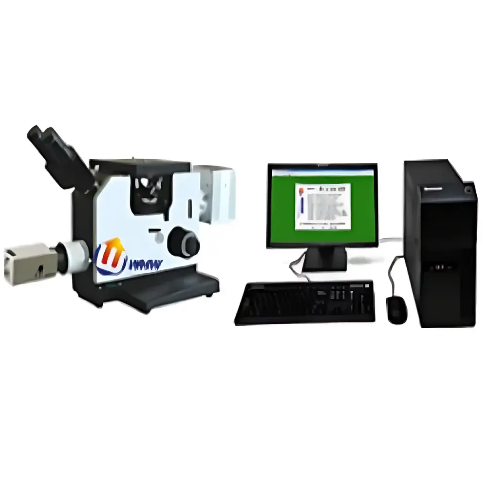
YANRUN AMM-8P Large-Scale R&D Inverted Horizontal Metallurgical Microscope
| Brand | YANRUN |
|---|---|
| Origin | Shanghai, China |
| Manufacturer Type | Direct Manufacturer |
| Product Category | Domestic |
| Model | AMM-8P Large-Scale R&D Inverted Horizontal Metallurgical Microscope |
| Mounting Configuration | Inverted |
| Image Analysis System | Not Standard (Optional Add-on) |
| Optical Total Magnification | 40×–400× |
| Optional Extended Magnification | 25×–1000× |
| Eyepieces | Wide-Field WF10X (Φ18 mm) |
| Objective Lenses | 4×, 10×, 20×, 40× |
| Coaxial Coarse/Fine Focus Travel | 15 mm / 0.002 mm (2 µm) per Division |
| Mechanical Stage | Dual-Layer, 180 × 155 mm Platform with 75 × 50 mm Travel Range |
| Illumination | Reflected Halogen Lamp, 6 V / 30 W, Continuously Adjustable Brightness |
| Power Supply | 220 V / 50 Hz |
Overview
The YANRUN AMM-8P is a large-scale, inverted horizontal metallurgical microscope engineered for rigorous materials science research, metallographic quality control, and failure analysis in industrial R&D laboratories. Unlike conventional upright configurations, its inverted optical layout positions the objective lenses beneath the specimen stage—enabling stable observation of large, heavy, or irregularly shaped metallic samples (e.g., castings, weldments, heat-treated plates, or machined components) without requiring vertical sample clamping or complex mounting fixtures. The system operates on Köhler illumination principles, utilizing reflected brightfield and optional darkfield contrast modes to resolve microstructural features—including grain boundaries, phase distributions, inclusion morphology, porosity, and interfacial reactions—with high axial resolution and consistent illumination uniformity across wide fields of view. Its robust mechanical architecture supports long-duration static imaging and sequential documentation under controlled environmental conditions, making it suitable for ISO/IEC 17025-accredited testing labs and GLP-compliant metallurgical characterization workflows.
Key Features
- Inverted horizontal design with 180 × 155 mm dual-layer mechanical stage and 75 × 50 mm travel range—optimized for oversized specimens up to 10 kg.
- Coaxial coarse/fine focusing mechanism with 15 mm total vertical travel and 0.002 mm (2 µm) fine-focus graduation for precise Z-axis positioning during depth-resolved microstructure assessment.
- Wide-field WF10X eyepieces (Φ18 mm field number) delivering ergonomic viewing at 45° inclination; compatible with optional Huygens 5X, wide-angle 10X (Φ20 mm), and 12.5X eyepieces for extended magnification flexibility.
- Standard parfocal, achromatic objective set (4×, 10×, 20×, 40×) with optional plan achromat upgrades including 2.5× (NA 0.07) and oil-immersion 100× (NA 0.85) for quantitative grain size analysis per ASTM E112.
- Reflected illumination module featuring a 6 V / 30 W halogen lamp with continuous intensity control—ensuring stable color temperature (~3200 K) and minimal thermal drift during extended observation sessions.
- Three-port trinocular head (standard 100:0 / 0:100 / 50:50 beam-splitting ratio) supporting simultaneous visual inspection, analog video output, and digital camera integration without optical path compromise.
Sample Compatibility & Compliance
The AMM-8P accommodates specimens up to 120 mm in height and 200 mm in width—ideal for cross-sectioned turbine blades, rolled steel slabs, additive-manufactured alloy coupons, and embedded metallographic mounts. Its low-profile stage clearance and reinforced base minimize vibration transmission, satisfying requirements for ASTM E3, ISO 643, and GB/T 13298 microstructural evaluation standards. While the base configuration does not include image analysis software, all optional imaging packages comply with FDA 21 CFR Part 11 data integrity guidelines when deployed with validated acquisition modules and audit-trail-enabled software (e.g., NIS-Elements or Olympus cellSens). The system’s electrical design conforms to IEC 61000-6-3 (EMC emission limits) and IEC 61010-1 (safety requirements for laboratory equipment).
Software & Data Management
Image capture and processing are supported via optional USB 3.0 or HDMI interfaces compatible with industry-standard machine vision cameras (e.g., Sony IMX series sensors). When integrated with third-party metrology platforms—such as Image-Pro Premier, Olympus Stream, or open-source Fiji/ImageJ—the microscope enables automated grain boundary tracing, phase fraction quantification, inclusion rating per ASTM E45, and hardness correlation mapping. All digital outputs adhere to TIFF 6.0 and DICOM-SR metadata conventions. Raw image files retain EXIF tags documenting objective ID, magnification, illumination intensity, and focus position—supporting traceable reporting in GMP-regulated environments. Audit logs, user authentication, and electronic signature capabilities are available through licensed software add-ons meeting ALCOA+ data governance criteria.
Applications
- Microstructural characterization of ferrous and non-ferrous alloys (aluminum, titanium, nickel-based superalloys) per ASTM E384 (microhardness correlation).
- Weld metal zone analysis—including fusion line delineation, heat-affected zone (HAZ) grain coarsening, and solidification cracking assessment.
- Failure analysis of fatigue fractures, stress corrosion cracking, and hydrogen embrittlement in aerospace and power generation components.
- Quality assurance of powder metallurgy parts, sintered ceramics, and composite matrix interfaces.
- Educational use in university metallurgy labs for hands-on training in specimen preparation, etching protocol validation, and stereological measurement techniques.
FAQ
Is the AMM-8P compatible with digital camera systems from major OEMs?
Yes—the trinocular port supports C-mount adapters for cameras from Canon, Nikon, Olympus, and Basler, with native driver support for Windows/Linux SDKs.
Can the system be upgraded to support polarized light or differential interference contrast (DIC)?
Polarization capability requires retrofitting with a strain-free objective set and rotating analyzer; DIC is not natively supported due to optical path constraints but may be implemented via external Nomarski prism modules.
What is the maximum specimen weight the stage can safely support?
The reinforced mechanical stage maintains positional stability for specimens up to 10 kg under static load, with dynamic damping effective up to 50 Hz ambient vibration frequency.
Does the halogen illumination system meet photostability requirements for time-lapse imaging?
While suitable for static documentation, prolonged exposure (>30 min) may induce thermal drift; LED illumination upgrade kits (5 W, 5500 K CCT) are available for enhanced stability and reduced IR emission.
Are calibration certificates provided for focus mechanisms and stage movement?
Factory calibration reports (traceable to NIM China) are included for fine-focus scale accuracy (±0.001 mm) and stage linear travel (±2 µm over full range); ISO/IEC 17025-certified recalibration services are offered biannually.


