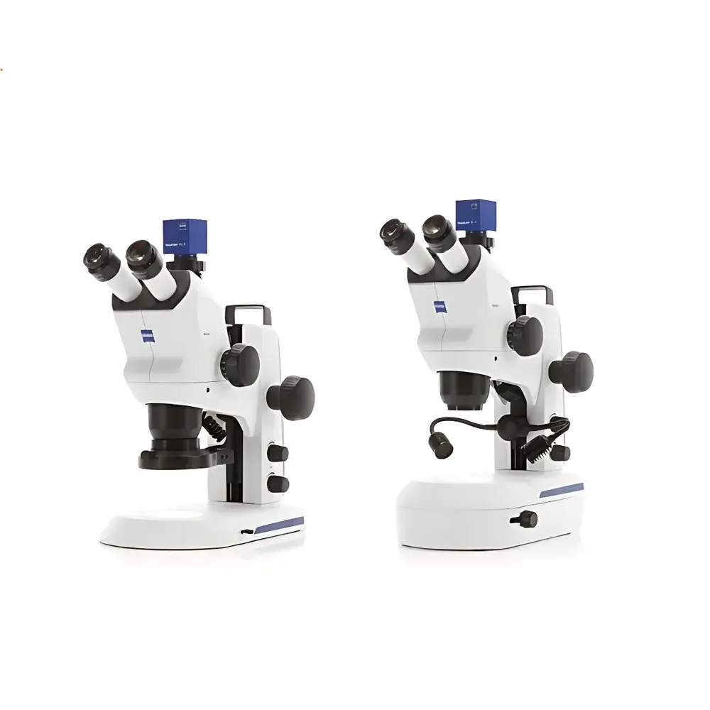
ZEISS Stemi 508 Stereo Microscope
| Brand | ZEISS |
|---|---|
| Origin | Germany |
| Model | Stemi 508 |
| Magnification Range | 6.3×–50× |
| Zoom Ratio | 8:1 |
| Field of View | up to 36 mm |
| Working Distance | 287 mm |
| Optical Correction | Apochromatic Zoom Body & Front Objective |
| Detented Zoom Positions | 10 |
| Illumination | Integrated LED System (Reflected, Transmitted, and Mixed Lighting) |
Overview
The ZEISS Stemi 508 is a high-performance stereo microscope engineered for routine inspection and documentation of macroscopic to mesoscopic specimens with pronounced topographic variation. Based on Greenough optical architecture—featuring dual, laterally separated objective paths—the system delivers natural depth perception, true stereoscopic imaging, and minimal parallax across the entire zoom range. Its apochromatically corrected zoom body and front objective eliminate chromatic and spherical aberrations across the full magnification spectrum (6.3×–50×), ensuring color fidelity and edge-to-edge sharpness critical for quantitative visual assessment and comparative morphological analysis. Designed for robust daily use in both laboratory and industrial environments, the Stemi 508 supports ergonomic observation workflows without compromising optical integrity or mechanical stability.
Key Features
- Apochromatic zoom optics with 8:1 zoom ratio and 10 precisely detented magnification positions—enabling repeatable, traceable settings for standardized inspection protocols.
- Maximum field of view of 36 mm at lowest magnification (6.3×), facilitating rapid survey of large-area samples such as PCB assemblies, tissue sections, or geological specimens.
- Extended working distance of 287 mm at 6.3×—accommodating bulky or multi-layered objects including microfluidic chips, battery cells, and live model organisms under controlled environmental conditions.
- Integrated modular LED illumination system offering three independent lighting modes: reflected (epi-illumination), transmitted (diascopic), and mixed—each individually adjustable in intensity and spatial configuration.
- Compact, space-efficient footprint optimized for benchtop integration in QC labs, R&D cleanrooms, and production line stations—compatible with optional motorized focus drives and digital camera adapters.
- Optomechanical design compliant with ISO 10934-1 (stereomicroscopes — nomenclature and metrological characteristics) and aligned with ZEISS’s long-standing adherence to DIN EN ISO 9001 manufacturing standards.
Sample Compatibility & Compliance
The Stemi 508 accommodates a broad spectrum of non-destructive, label-free sample types—from live zebrafish embryos imaged under oblique transmitted illumination to solder joints inspected under quarter-ring epi-LED illumination. Its large working distance and versatile illumination enable stable observation of delicate biological preparations (e.g., parasitological mounts, insect dissections), precision-manufactured components (e.g., MEMS devices, watch gears), and heterogeneous geological thin sections. The system meets requirements for GLP-compliant documentation workflows when paired with ZEISS ZEN Core software, supporting audit trails, user access control, and metadata embedding per FDA 21 CFR Part 11 guidelines where applicable.
Software & Data Management
When coupled with ZEISS ZEN Core imaging software (sold separately), the Stemi 508 enables structured image acquisition, annotation, measurement (length, angle, area), and report generation. ZEN Core supports DICOM export, TIFF/OME-TIFF archival formats, and batch processing for multi-field montaging. Measurement data can be exported to CSV or Excel for statistical analysis in QA/QC frameworks. Software validation packages are available for regulated industries requiring IQ/OQ/PQ documentation and electronic record integrity per ISO/IEC 17025 and ASTM E2919-22 (Standard Guide for Digital Imaging in Microscopy).
Applications
- Electronics & Semiconductor: Visual inspection of PCB surface defects, solder joint integrity, wire bond alignment, and conformal coating uniformity.
- Automotive & Aerospace: Fracture surface analysis, weld seam profiling, gasket interface evaluation, and composite material delamination detection.
- Battery Manufacturing: Electrode surface morphology assessment, separator pore structure screening, and tab weld dimensional verification.
- Life Sciences: Embryonic development staging (e.g., zebrafish, Drosophila), plant organogenesis studies, entomological taxonomy, and marine specimen curation.
- Forensics & Document Examination: Handwriting comparison, ink differentiation, latent print enhancement, and fiber morphology analysis under variable contrast illumination.
- Geosciences & Paleontology: Fossil surface texture mapping, mineral grain boundary delineation, and thin-section petrographic correlation.
FAQ
Is the Stemi 508 compatible with third-party digital cameras?
Yes—via C-mount or F-mount adapters (available as optional accessories), provided the sensor size and flange distance align with ZEISS mechanical interface specifications.
Can the LED illumination be calibrated for photometric consistency across users?
While the integrated LEDs are not factory-calibrated to NIST-traceable radiometric standards, intensity output remains stable over time and can be normalized using ZEN Core’s reference image function for longitudinal comparison.
Does the Stemi 508 support motorized zoom or focus functions?
Motorization is not built-in but can be added via the optional ZEISS Motorized Focus Drive (MFD) and compatible controllers, enabling programmable Z-stacking and automated multi-position imaging.
What is the recommended maintenance interval for optical alignment verification?
ZEISS recommends annual optomechanical verification by certified service personnel; interim user-level checks (e.g., reticle calibration, coaxial alignment test) are outlined in the operator manual.
Are replacement objectives or auxiliary lenses available for extended magnification ranges?
Yes—ZEISS offers supplementary front objectives (e.g., 0.5×, 1.6×, 2.0×) and eyepiece options (10×, 15×, 20× widefield) to extend effective magnification from 3.15× to 100× while maintaining resolution and depth of field integrity.


