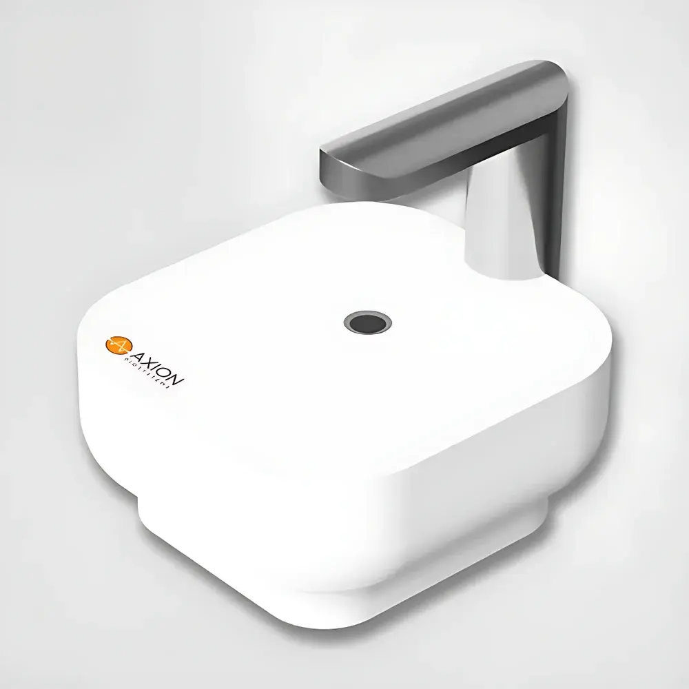

Axion BioSystems Lux3 In-Box Brightfield and Fluorescence Live-Cell Imaging Analysis System
| Brand | Axion BioSystems |
|---|---|
| Origin | Netherlands |
| Manufacturer | Axion BioSystems |
| Product Type | Imported Instrument |
| Model | Lux3 |
| Temperature Control Range | 5–40 °C |
| Humidity Control Range | 20–95% RH |
Overview
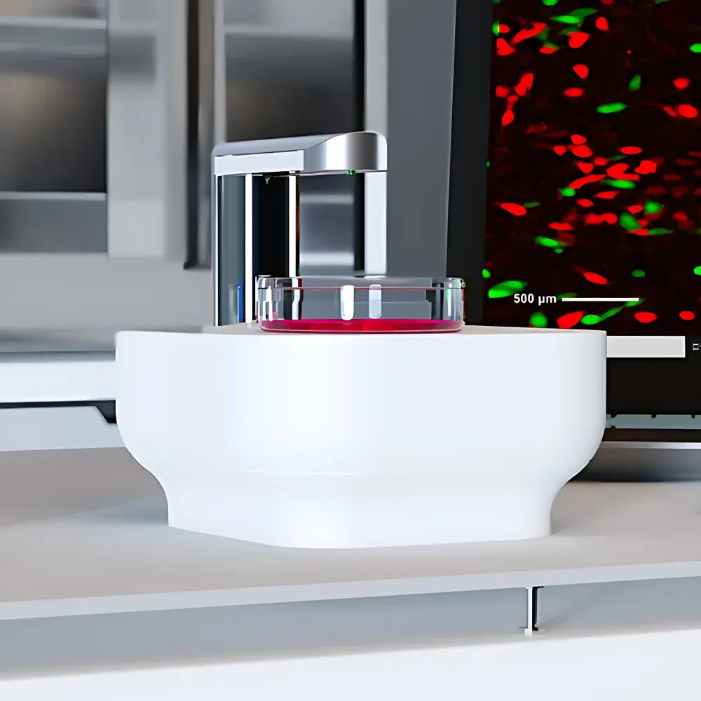
The Axion BioSystems Lux3 In-Box Brightfield and Fluorescence Live-Cell Imaging Analysis System is an engineered solution for non-invasive, long-term kinetic monitoring of adherent mammalian cells directly inside standard CO2 incubators. Built upon a compact optical architecture optimized for sterile, humidified, temperature-controlled environments, the Lux3 employs inverted brightfield and dual-channel (green/red) epifluorescence microscopy principles—leveraging high-efficiency LED excitation and a precision-corrected CMOS sensor—to capture time-lapse image sequences without disturbing culture conditions. Unlike conventional benchtop imagers requiring sample removal, the Lux3 eliminates thermal and pH transients associated with incubator door opening, thereby preserving physiological relevance across experiments lasting hours to weeks. Its design complies with fundamental requirements for live-cell assay integrity: stable focus maintenance under thermal drift, minimal phototoxicity via intensity-tunable illumination, and hardware-level synchronization between exposure timing and environmental stability.
Key Features
- In-box deployment: Fully functional within standard CO2 incubators (5–40 °C, 20–95% RH); no external optics or vibration-sensitive components.
- Dual-modality imaging: Simultaneous brightfield and fluorescence (ex: 470/525 nm and 535/620 nm) acquisition with automatic channel switching and exposure optimization.
- Compact form factor: Footprint under 180 × 180 mm; height ≤ 120 mm—designed to fit beneath standard shelf configurations without compromising growth space.
- Automated time-lapse operation: Programmable acquisition intervals (1 min to 24 h), multi-position scanning per well, and Z-stack capability (up to 5 planes).
- Zero-maintenance optics: Fixed-focus lens system with factory-calibrated working distance; no user alignment or periodic recalibration required.
- Remote accessibility: Real-time image streaming and experiment control via secure HTTPS connection to CytoSMART Cloud; compatible with Windows, macOS, and Linux clients.
Sample Compatibility & Compliance
The Lux3 accommodates all standard transparent, flat-bottom cell culture vessels with total height ≤ 55 mm—including 6–384-well plates, Petri dishes (35–150 mm), T-flasks (T25 to T225), and microfluidic chips with optical-grade PDMS or glass substrates. Its mechanical stage supports both manual positioning and automated XY mapping across multi-well formats. From a regulatory standpoint, the system supports GLP- and GMP-aligned workflows through audit-trail-enabled software logging (user actions, timestamps, parameter changes), raw data export in TIFF/OME-TIFF format, and compatibility with 21 CFR Part 11-compliant electronic signature add-ons when deployed with enterprise CytoSMART Suite licenses. It meets IEC 61000-6-3 (EMC emission) and IEC 61010-1 (safety) standards for laboratory equipment operating in Class II biological safety environments.
Software & Data Management
Acquired images are transmitted automatically to the CytoSMART Cloud platform, where they undergo on-server processing using validated, ISO/IEC 17025-aligned image analysis algorithms. Users may select from modular applications including confluence quantification (brightfield and fluorescence), scratch wound healing kinetics, colony formation analysis (CFU counting with size/threshold filtering), and fluorescent object segmentation (nuclei/cytoplasm labeling). All modules generate CSV- and PDF-formatted reports compliant with internal SOP templates. Raw datasets remain fully accessible for reprocessing in third-party platforms such as ImageJ/Fiji, MATLAB, or Python-based scikit-image pipelines. Version-controlled software updates follow a documented change control process, with release notes archived per ISO 9001 requirements.
Applications
- Cell proliferation kinetics: Quantitative confluence tracking across multiple passages; detection of subtle growth inhibition or hyperproliferation phenotypes.
- Cytotoxicity profiling: Real-time morphological assessment (blebbing, detachment, granularity) combined with fluorescent viability dyes (e.g., Calcein-AM/PI) for EC50 determination.
- Immunooncology assays: Monitoring CAR-T or NK-cell-mediated target lysis dynamics in co-culture models using dual-label fluorescence (target GFP + effector RFP).
- Migratory behavior analysis: Automated wound closure measurement in monolayer scratch assays, including velocity vector field derivation and edge roughness metrics.
- Stem cell differentiation monitoring: Morphometric classification of colony heterogeneity during spontaneous or induced differentiation protocols.
FAQ
How does the Lux3 acquire images without disrupting incubator conditions?
The system uses an inverted optical path: LED illumination is delivered from above the sample, while imaging occurs through the bottom of the vessel via a fixed-focus lens positioned beneath the stage—eliminating the need for top-access optics or mechanical movement inside the chamber.
Which image analysis modules are available off-the-shelf?
Pre-validated modules include Brightfield/Fluorescence Confluence Analysis, Scratch Wound Healing Quantification, Colony Formation Assay, and Fluorescent Object Counting. Custom algorithm development services are available under NDA.
Can the Lux3 be integrated into existing LIMS or ELN systems?
Yes—via RESTful API access to CytoSMART Cloud, enabling bidirectional metadata exchange (sample IDs, protocol parameters, results) with major laboratory informatics platforms supporting JSON/XML payloads.
What is the maximum supported vessel height?
55 mm from stage surface to lower edge of illumination module—compatible with stacked multi-layer plates and tall flasks when placed on elevated staging inserts (optional).
Is calibration required before each experiment?
No. The optical path is factory-aligned and thermally stabilized; only initial setup validation (using provided reference slide) is recommended per ISO 21531:2020 guidelines for imaging system verification.


