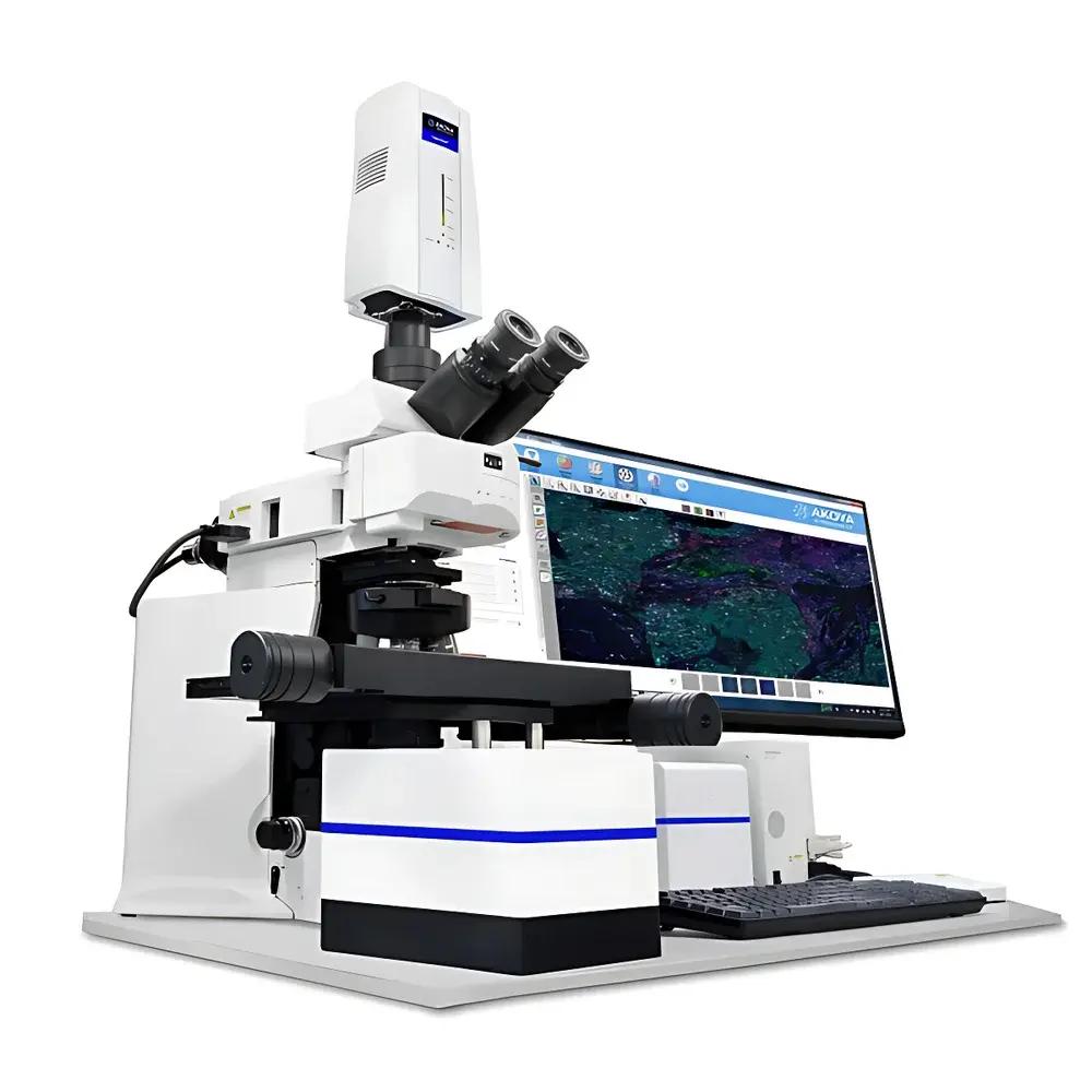
Akoya Vectra 3 Automated Multispectral Tissue Imaging and Quantitative Analysis System
| Brand | Akoya |
|---|---|
| Origin | USA |
| Model | Vectra 3 |
| Price Range | USD $420,000 – $700,000 |
| Application Domain | Multiplex Immunofluorescence (mIF) and Hematoxylin & Eosin (H&E) Digital Pathology |
| Imaging Modality | High-Resolution Multispectral Fluorescence Microscopy |
| Throughput | Up to 6 Tissue Sections per Run |
| Software Platform | inForm Advanced Image Analysis Suite |
Overview
The Akoya Vectra 3 Automated Multispectral Tissue Imaging and Quantitative Analysis System is an integrated digital pathology platform engineered for reproducible, high-fidelity spatial profiling of formalin-fixed paraffin-embedded (FFPE) and frozen tissue sections. Built upon multispectral unmixing technology, the system captures full-spectrum fluorescence emission data across multiple excitation wavelengths—enabling precise separation of spectrally overlapping fluorophores without reliance on sequential staining or physical filter wheels. Unlike conventional widefield fluorescence microscopes, the Vectra 3 employs a tunable liquid crystal tunable filter (LCTF) coupled with a scientific-grade monochrome CCD camera to acquire 31 discrete spectral bands per pixel, followed by linear unmixing algorithms that resolve contributions from individual biomarkers—even those with <15 nm spectral separation. This principle supports quantitative co-expression analysis, cell phenotyping, and spatial context mapping at single-cell resolution within intact tissue architecture.
Key Features
- Fully automated slide loading and autofocus: accommodates up to six standard glass slides (1″ × 3″) with barcode recognition and positional calibration for walk-away operation.
- Multispectral imaging engine: acquires 31-band spectral stacks at 20× and 40× magnifications (0.5 µm/pixel at 40×), supporting both brightfield (H&E) and multiplex immunofluorescence (up to 6-plex in a single run).
- Integrated spectral library generation: users can define custom reference spectra from control tissues or commercial standards, enabling robust unmixing across diverse antibody-fluorophore conjugates (e.g., Opal™ dyes, Alexa Fluor®, Cy® dyes).
- Hardware-based background subtraction and autofluorescence removal: real-time correction using tissue-specific spectral signatures, minimizing false-positive detection in lipofuscin-rich or collagen-dense regions.
- Modular optical path: compatible with standard microscope objectives (Nikon CFI Plan Apo series) and optional polarization filters for birefringence-assisted collagen segmentation.
Sample Compatibility & Compliance
The Vectra 3 accepts standard histopathology slides prepared via routine H&E, IHC, or multiplex IF protocols—including antigen retrieval, blocking, and tyramide signal amplification (TSA)-based workflows. It supports both manual and automated staining platforms (e.g., Leica Bond RX, Ventana Discovery Ultra). All image acquisition and analysis workflows comply with CAP/CLIA documentation requirements for clinical research use. The inForm software meets FDA 21 CFR Part 11 criteria for electronic records and signatures when deployed with validated IT infrastructure, including audit trail logging, user access controls, and versioned analysis templates. Data export formats (e.g., TIFF, OME-TIFF, CSV) are compatible with downstream tools such as HALO®, Visiopharm®, and QuPath for cross-platform validation.
Software & Data Management
The inForm software suite provides end-to-end computational pathology support—from spectral unmixing and tissue segmentation to cell classification and spatial statistics. Its machine learning–enabled tissue classifier distinguishes stroma, tumor, necrosis, and lymphoid aggregates with >92% concordance against expert pathologist annotations (per internal validation studies). Cell-level phenotyping uses hierarchical gating logic based on intensity thresholds, morphometric features (nuclear area, cytoplasmic ratio, shape descriptors), and proximity metrics (e.g., distance to nearest CD8+ T cell). All analysis parameters are saved as reusable protocols with metadata tagging; raw spectral cubes and processed layers are stored in vendor-neutral formats to ensure long-term archival integrity and interoperability with institutional PACS or LIMS environments.
Applications
- Translational immuno-oncology: quantifying immune cell infiltration patterns (e.g., PD-1/PD-L1 co-expression, Treg/CD8+ ratios) in tumor microenvironments across clinical trial cohorts.
- Biomarker discovery and assay development: validating novel antibody panels for diagnostic or prognostic utility in solid tumors and hematologic malignancies.
- Neurodegenerative disease research: mapping amyloid-beta plaque burden alongside microglial activation states in postmortem brain sections.
- Toxicologic pathology: assessing drug-induced tissue injury through multiplexed detection of apoptosis markers (cleaved caspase-3), proliferation indices (Ki-67), and lineage-specific antigens.
- Regulatory submission support: generating analytically validated image-derived endpoints aligned with ICH M10 and ISAC guidelines for quantitative digital pathology endpoints.
FAQ
What sample preparation methods are validated for use with the Vectra 3?
Standard FFPE sectioning (4–5 µm), deparaffinization, epitope retrieval (heat-induced or enzymatic), and Opal™-based multiplex IF protocols are fully supported. Frozen sections (5–10 µm) and cytospin preparations are also compatible with protocol optimization.
Can the Vectra 3 perform true 7-plex or higher multiplexing?
Yes—through iterative staining cycles (e.g., Opal™ TSA with stripping), the platform enables serial multiplexing beyond six markers. Spectral unmixing remains valid provided reference spectra are reacquired after each round.
Is remote system monitoring available?
The Vectra 3 supports secure SSH-based remote diagnostics and log review via Akoya’s authorized service portal. Routine maintenance alerts and instrument health metrics are accessible through the embedded web interface.
How is analytical reproducibility ensured across instruments and sites?
Akoya provides factory-calibrated spectral reference standards and inter-laboratory harmonization kits. In addition, the inForm software includes batch correction modules trained on multi-site validation datasets to minimize inter-instrument variance in intensity normalization.
Does the system support whole-slide imaging (WSI) export?
Yes—unmixed biomarker maps and composite overlays can be exported as pyramid TIFFs compliant with ASAM WSI standards, enabling integration into enterprise digital pathology viewers such as Philips IntelliSite or 3DHISTECH CaseViewer.
Related Products


