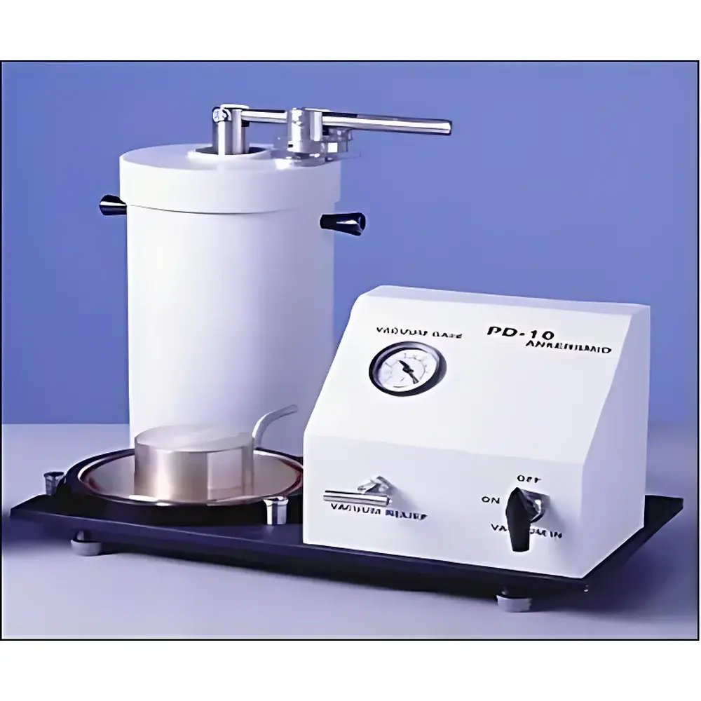
Ankersmid PD-10 Microscope Slide Dry Powder Dispersion Analyzer
| Brand | Ankersmid |
|---|---|
| Origin | Netherlands |
| Model | PD-10 |
| Instrument Type | Pneumatic Impact Dispersion System |
| Sample Handling Principle | Vacuum-Induced Turbulent Shear Dispersal |
| Compliance Context | Designed for ISO 13322-1 (particle morphology by image analysis), ASTM E2557 (dry powder dispersion for microscopy), GLP-compliant sample preparation workflows |
Overview
The Ankersmid PD-10 Microscope Slide Dry Powder Dispersion Analyzer is an engineered solution for reproducible, morphology-preserving dry powder dispersion prior to optical or electron microscopic analysis. Unlike mechanical grinding, sonication, or manual deposition methods—which risk particle fracture, agglomeration, or non-uniform monolayer formation—the PD-10 employs a controlled pneumatic impact principle grounded in fluid dynamics. Upon actuation, the system rapidly evacuates the dispersion chamber to sub-atmospheric pressure; triggering the vacuum seal release generates a transient, high-Reynolds-number air jet. This turbulent flow subjects aggregated particles to instantaneous shear stresses sufficient to overcome van der Waals and electrostatic inter-particle forces—without introducing thermal degradation or mechanical attrition. Concurrently, gravity-driven sedimentation occurs under minimized aerodynamic drag due to the low-pressure environment, enabling near-synchronous settling velocities across particle size distributions (typically 0.5 µm to 200 µm). The result is a statistically representative, non-overlapping monolayer on standard 25 × 75 mm microscope slides—critical for accurate particle sizing, shape factor quantification (e.g., circularity, aspect ratio), and automated image-based classification.
Key Features
- Pneumatic impact dispersion mechanism eliminates operator-dependent variability in dry powder deposition
- Single-use or cleanable stainless-steel dispersion head with rapid-access design—full disassembly and cleaning completed in under 60 seconds using ethanol or IPA
- Compatible with hazardous, hygroscopic, or toxic powders via optional sealed glove-box integration (ISO Class 5 laminar flow enclosure recommended)
- No moving parts in contact with sample—prevents cross-contamination and wear-induced calibration drift
- Calibration traceable to NIST-traceable reference powders (e.g., SRM 1980) for inter-laboratory comparability
- Designed for integration with automated stage microscopes and third-party image analysis platforms (including EyeTech software suite)
Sample Compatibility & Compliance
The PD-10 accommodates a broad spectrum of dry particulate materials: low-density fumed silica, high-density metal oxides (e.g., TiO₂, Fe₃O₄), pharmaceutical excipients (lactose, microcrystalline cellulose), catalyst supports (alumina, zeolites), and nanoscale carbon blacks. It meets functional requirements outlined in ISO 13322-1:2020 for static image analysis of particle morphology, and supports compliance with USP , EP 2.9.31, and FDA guidance on analytical method validation for solid dosage forms. When used within validated SOPs—including defined vacuum level (–85 kPa ± 2 kPa), actuation dwell time (120 ms), and ambient RH control (<40%)—the system delivers RSD <3.2% for particle count per field-of-view across five independent operators (n = 30 slides, 500× magnification).
Software & Data Management
While the PD-10 operates as a standalone hardware module, its output is optimized for digital microscopy workflows. Slides prepared with the PD-10 are directly compatible with EyeTech’s GMP-auditable image analysis software (v5.4+), which provides 21 CFR Part 11–compliant audit trails, electronic signatures, and metadata embedding (including instrument ID, date/time stamp, operator ID, and dispersion parameters). Raw slide images retain EXIF tags with acquisition settings; processed data exports to CSV, PDF, and XML formats for LIMS ingestion. No proprietary drivers or dongles are required—standard USB 3.0 or GigE Vision interfaces suffice for camera coupling.
Applications
- Pharmaceutical quality control: verification of milled API crystallinity and agglomerate disruption post-milling
- Advanced materials R&D: characterization of sintering behavior predictors (e.g., specific surface area proxies via projected area distribution)
- Environmental monitoring: identification and quantification of airborne particulate matter (PM₁₀, PM₂.₅) collected on filters or slides
- Geological sample prep: dispersion of clay minerals and silicates without altering platy morphology
- Regulatory submissions: generation of morphology evidence packages compliant with ICH Q5A(R2) for biotechnology-derived products
FAQ
Does the PD-10 require compressed gas or external vacuum pumps?
No. The PD-10 integrates a built-in diaphragm vacuum pump capable of achieving –85 kPa absolute pressure; no auxiliary utilities are needed.
Can it be used with conductive or electrostatic-sensitive powders?
Yes. The dispersion chamber includes grounded stainless-steel components and optional anti-static coating; relative humidity control below 40% further mitigates charge accumulation.
Is routine calibration required?
Annual verification against NIST-traceable reference powders is recommended; no field recalibration is necessary between uses due to solid-state actuation mechanics.
What slide types are supported?
Standard plain glass, frosted-end, or conductive ITO-coated slides (max thickness 1.2 mm); compatibility confirmed with Leica, Zeiss, and Olympus stage adapters.
How does PD-10 compare to ultrasonic nebulization for microscopy prep?
Ultrasonic methods induce cavitation-induced particle fragmentation and solvent-mediated reagglomeration; PD-10 preserves native morphology via dry, shear-dominated dispersion—validated by SEM cross-correlation studies.


