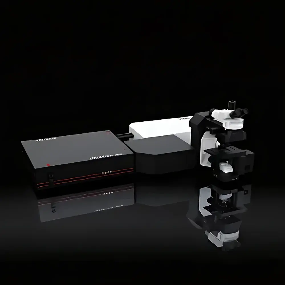
VibroniX UltraView MK-III Stimulated Raman Scattering (SRS) Microscopic Imaging System
| Brand | VibroniX |
|---|---|
| Origin | Jiangsu, China |
| Manufacturer Type | Authorized Distributor |
| Country of Origin | China |
| Model | UltraView MK-III |
| Instrument Type | Stimulated Raman Scattering (SRS) Microscope |
| Spectral Coverage | 500–3200 cm⁻¹ |
| Spatial Resolution | 350 nm |
| Imaging Speed | 10 FPS |
Overview
The VibroniX UltraView MK-III is a high-performance, multimodal coherent Raman microscopic imaging system engineered for label-free, chemically specific visualization of biological and material samples at subcellular resolution. It operates on the physical principle of stimulated Raman scattering (SRS), a nonlinear optical process in which two synchronized femtosecond laser beams—a pump beam (ωp) and a Stokes beam (ωs)—interact within the sample to resonantly excite molecular vibrational modes when their frequency difference (ωp − ωs) matches a target Raman shift. This results in coherent, background-free signal amplification proportional to molecular concentration—enabling quantitative, video-rate chemical mapping without exogenous dyes or fixation artifacts. Complementing SRS, the system natively supports coherent anti-Stokes Raman scattering (CARS) and two-photon excited fluorescence (TPEF), allowing simultaneous acquisition of intrinsic molecular contrast and genetically encoded or synthetic fluorophore signals. Its integrated architecture leverages ultrafast fiber-based laser sources, high-numerical-aperture water- or oil-immersion objectives, and lock-in detection electronics to achieve diffraction-limited spatial resolution (350 nm), broad spectral coverage (500–3200 cm⁻¹), and real-time volumetric imaging (up to 10 frames per second). Designed for rigorous life science research, the UltraView MK-III meets the operational requirements of live-cell dynamics, thick-tissue penetration, and longitudinal in vivo studies under physiologically relevant conditions.
Key Features
- Label-free chemical specificity: Direct detection of endogenous biomolecules—including lipids (C–H stretch at ~2845 cm⁻¹), proteins (amide I at ~1650 cm⁻¹), nucleic acids (C≡N at ~2230 cm⁻¹), and pharmaceuticals—via tunable vibrational resonance, eliminating labeling-induced perturbations.
- Video-rate imaging capability: Achieves 10 FPS full-frame acquisition with sub-millisecond pixel dwell time, enabling real-time tracking of metabolic flux (e.g., D-glucose uptake), membrane trafficking, and neural activity without motion blur.
- Sub-diffraction spatial fidelity: Delivers lateral resolution of 350 nm using high-NA objectives and optimized beam delivery, resolving subcellular organelles such as lipid droplets, mitochondria, and myelin sheaths in native tissue context.
- Native multimodal integration: Seamless co-registration of SRS/CARS chemical contrast with TPEF structural/functional signals and optional SHG collagen imaging—all acquired under identical optical path and coordinate frame.
- Optical sectioning and 3D reconstruction: Supports Z-stack acquisition with axial resolution < 1 µm, enabling volumetric chemical tomography of organoids, brain slices, and skin equivalents without physical sectioning.
- Bio-compatible illumination strategy: Employs near-infrared femtosecond pulses (typically 780–1040 nm) with peak power management and adaptive exposure control to minimize phototoxicity during extended live-sample observation.
- Automated workflow engine: Includes motorized stage control, autofocus routines, multi-position time-lapse sequencing, and spectral tuning presets for rapid transition between molecular targets (e.g., CH₂ vs. CD₂ bonds).
Sample Compatibility & Compliance
The UltraView MK-III accommodates diverse biological specimens including adherent and suspension mammalian cells, primary neuronal cultures, ex vivo tissue sections (up to 500 µm thickness), intact zebrafish embryos, murine brain slices, human-derived organoids (cerebral, intestinal, tumor), plant tissues (stem cross-sections, stomatal guard cells), and environmental microplastic particles. Sample mounting follows standard protocols for aqueous immersion or coverslip-based preparations; no dehydration, embedding, or staining is required. The system complies with ISO 13485 design controls for medical device-related research instrumentation and supports audit-ready data integrity through timestamped metadata logging. While not a diagnostic device per FDA 21 CFR Part 820, its data output conforms to GLP/GMP-aligned documentation standards—including electronic lab notebook (ELN) export, raw spectral cube storage (HDF5), and traceable instrument parameter records—facilitating regulatory submissions in preclinical development workflows.
Software & Data Management
Control and analysis are unified within the proprietary UltraView Acquisition Suite, a Windows-based application built on modular C++/Python architecture. Core modules include Laser Tuning Manager (for precise wavenumber selection across 500–3200 cm⁻¹), Real-Time SRS Engine (with hardware-synchronized lock-in demodulation), Multichannel Registration Tool (for pixel-accurate SRS/TPEF/SHG overlay), and Quantitative Spectral Analyzer (supporting peak fitting, ratio imaging, and PCA-based unsupervised clustering). All datasets are saved in vendor-neutral HDF5 format with embedded metadata compliant with the OME-NGFF specification. Batch processing pipelines support automated background subtraction, drift correction, and 3D deconvolution. For regulatory environments, the software optionally enables 21 CFR Part 11-compliant user authentication, electronic signatures, and immutable audit trails—fully configurable to institutional IT security policies.
Applications
- Cellular metabolism: Dynamic tracking of deuterated metabolites (e.g., D-glucose, D-leucine) in live cancer cells and stem cell derivatives.
- Oncology research: Intraoperative margin assessment via lipid/protein ratio mapping in glioblastoma and prostate carcinoma biopsies.
- Neuroscience: Myelin integrity quantification in transgenic mouse models of ALS and MS; synaptic protein synthesis imaging in hippocampal organotypic slices.
- Synthetic biology: Spatial profiling of engineered metabolic pathways in bacterial colonies and yeast spheroids.
- Pharmaceutical development: Intracellular drug localization and lysosomal accumulation kinetics (e.g., imatinib, nilotinib) in 3D tumor models.
- Organoid science: 4D metabolic cartography of cerebral and cardiac organoids during differentiation and disease modeling.
- Environmental analysis: Identification and morphological characterization of polyethylene, PET, and nylon microplastics in aqueous and cellular matrices.
- Plant physiology: In situ cellulose crystallinity mapping and stomatal aperture dynamics under abiotic stress.
FAQ
What laser sources are integrated into the UltraView MK-III?
The system employs turnkey, maintenance-free femtosecond fiber lasers: one tunable optical parametric oscillator (OPO) for the Stokes beam (680–950 nm) and a fixed-wavelength Yb-fiber amplifier for the pump beam (1040 nm), enabling continuous spectral coverage from 500 to 3200 cm⁻¹.
Can the system perform hyperspectral SRS imaging?
Yes—the UltraView MK-III supports step-scan and continuous-tuning hyperspectral acquisition modes, generating full Raman spectra per pixel for multivariate analysis and untargeted chemical discovery.
Is TPEF imaging fully synchronized with SRS acquisition?
Yes—both modalities share the same pulsed excitation source and scanning galvanometer platform, ensuring temporal and spatial registration within ±50 ns and ±50 nm, respectively.
What objective lenses are supported?
Standard configurations include 25×/1.05 NA water-dipping, 40×/1.15 NA water-immersion, and 60×/1.28 NA silicone-oil objectives; custom lens interfaces are available upon request.
Does the system meet international calibration standards?
All optical paths undergo factory calibration against NIST-traceable Raman standards (e.g., cyclohexane, silicon); spectral accuracy is certified to ±1.5 cm⁻¹ over the full range, and spatial calibration is verified using USAF 1951 resolution targets.


