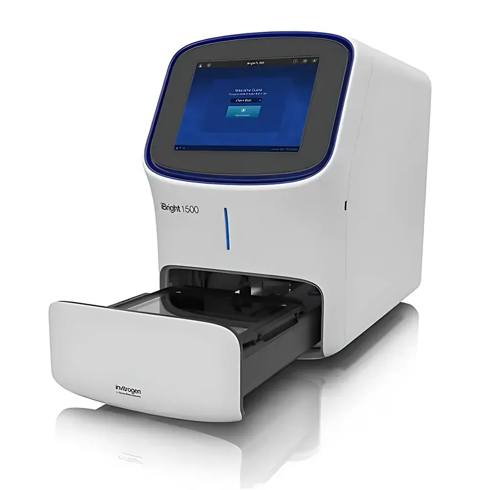
Thermo Fisher iBright FL1500 Gel Imaging System
| Brand | Thermo Fisher |
|---|---|
| Origin | USA |
| Model | A44241CFR |
| Instrument Type | Multicolor Fluorescent Gel Imaging System |
| CCD Camera | 9.1 MP Cooled CCD |
| Dimensions (W×D×H) | 68 × 38 × 60 cm |
| Field of View | 22.5 × 18.0 cm |
| Excitation Source | Green LED Transilluminator |
| Fluorescence Channels | 5 |
| Interface | 12.1" Capacitive Touchscreen |
| Connectivity | Ethernet, Wi-Fi (optional), USB, Cloud Export (Thermo Fisher Connect) |
Overview
The Thermo Fisher iBright FL1500 Gel Imaging System is a high-performance, integrated platform engineered for quantitative imaging of nucleic acid gels, protein gels, and Western blots across fluorescence, chemiluminescence, and colorimetric detection modalities. Built upon a cooled 9.1-megapixel CCD architecture with thermoelectric cooling to −25 °C, the system delivers low-noise acquisition essential for detecting weak signals in low-abundance targets—particularly critical in multiplexed fluorescent Western blotting and sensitive DNA visualization. Its optical design incorporates a precision green LED transilluminator (peak emission ~525 nm), optimized for SYBR Safe, SYBR Green I/II, ethidium bromide, and other common nucleic acid dyes—eliminating UV exposure risks while maintaining high excitation efficiency. The system operates on a fixed-optics principle with motorized lens positioning and auto-alignment algorithms, ensuring consistent magnification and spatial fidelity across repeated acquisitions. Designed for compliance-ready laboratories, it supports audit-trail–enabled workflows aligned with GLP and GMP documentation requirements.
Key Features
- Cooled 9.1 MP CCD sensor with −25 °C thermoelectric cooling for enhanced signal-to-noise ratio and extended integration times
- Five independently configurable fluorescence excitation/emission channels enabling simultaneous detection of up to four proteins per blot using spectrally distinct fluorophores (e.g., IRDye 680RD, Alexa Fluor 488, Cy3, Cy5)
- Smart Exposure technology that automatically calculates optimal exposure duration based on real-time preview histogram analysis—reducing trial-and-error iterations
- High Dynamic Range (HDR) imaging mode: intelligently fuses multiple exposures into a single linear image with >4-log dynamic range, preserving both saturated and subthreshold pixel intensities without clipping
- 12.1-inch capacitive touchscreen interface with intuitive icon-driven workflow navigation; no external PC required for basic acquisition and analysis
- Automated sample handling: motorized stage rotation (0°/90°/180°/270°), auto-zoom, and contrast-optimized autofocus ensure reproducible framing and focus across diverse sample formats
- 22.5 × 18.0 cm field of view accommodates up to four mini-blots (e.g., 8 × 10 cm) or two midi-blots (e.g., 13.5 × 16 cm) in a single capture—increasing throughput without compromising resolution
- Green LED transilluminator with uniform intensity distribution (±5% across FOV) and adjustable intensity control for consistent nucleic acid gel excitation
Sample Compatibility & Compliance
The iBright FL1500 supports a broad range of life science sample formats including polyacrylamide and agarose gels, nitrocellulose and PVDF membranes, microtiter plates (6–96-well), and colony filters. It is validated for use with standard chemiluminescent substrates (e.g., SuperSignal West Pico/Femto), fluorescent dyes (SYBR series, GelRed, Deep Purple), and colorimetric reagents (Coomassie Brilliant Blue, Ponceau S). From a regulatory perspective, the system’s firmware and embedded software comply with FDA 21 CFR Part 11 requirements when used with Thermo Fisher Connect cloud services—including electronic signatures, role-based access control, and immutable audit trails. Data files are saved in vendor-neutral TIFF and multi-page PDF formats, facilitating long-term archival and third-party analysis interoperability.
Software & Data Management
Acquisition and primary analysis are performed via the onboard iBright OS—a Linux-based, locked-down operating system optimized for stability and security. Quantitative tools include background subtraction, lane/band detection, molecular weight estimation, relative density calculation, and normalization against loading controls (e.g., β-actin, GAPDH). Raw image data can be exported directly to Thermo Fisher Connect, where users access iBright Analysis Software (desktop and web versions) for advanced features such as multiplex band deconvolution, statistical comparison across replicates, and automated report generation compliant with ISO/IEC 17025 documentation standards. All export operations—whether via USB drive, Ethernet SMB share, or secure HTTPS upload—preserve EXIF metadata including exposure time, gain, lens position, and calibration timestamp.
Applications
The iBright FL1500 is routinely deployed in academic core facilities, biopharmaceutical QC labs, and contract research organizations for applications including: validation of CRISPR editing efficiency via fluorescent genotyping gels; quantification of phosphorylated signaling proteins in dose-response studies; comparative analysis of protein expression across tissue lysates in biomarker discovery; verification of RNA integrity post-extraction using ribosomal RNA band ratios; and colony-forming unit (CFU) enumeration in antibiotic susceptibility assays. Its ability to resolve co-migrating bands with differential fluorophore labeling enables rigorous validation of antibody specificity and cross-reactivity profiles—supporting ICH Q5E comparability assessments during biosimilar development.
FAQ
Does the iBright FL1500 support time-lapse chemiluminescent imaging?
Yes—the system allows programmable sequential acquisition with user-defined intervals (1 sec to 24 hrs), ideal for kinetic substrate reactions such as luminol-based detection.
Can I perform spectral unmixing of overlapping fluorophores?
Spectral unmixing is not performed onboard; however, multi-channel TIFF stacks retain full spectral metadata and are compatible with third-party unmixing tools (e.g., ImageJ/Fiji plugins, MATLAB-based deconvolution scripts).
Is calibration traceable to NIST standards?
Yes—each instrument ships with a factory-calibrated reference slide certified for pixel intensity linearity and uniformity, with calibration data traceable to NIST-traceable photometric standards.
What is the maximum recommended exposure time for chemiluminescent detection?
Up to 60 minutes at full sensor gain; extended integrations are supported with active cooling maintaining thermal noise below 0.5 e⁻/pixel/sec.
Does the system meet CE and FCC regulatory requirements?
Yes—the A44241CFR model carries CE marking (EN 61010-1, EN 55011) and FCC Class B certification for electromagnetic compatibility in laboratory environments.


