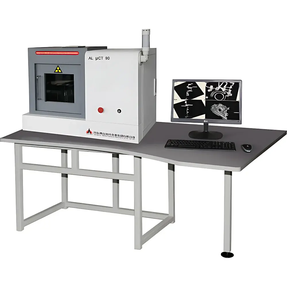
Aolong Desktop Micro-CT System (μCT)
| Brand | Aolong |
|---|---|
| Origin | Liaoning, China |
| Manufacturer Type | Original Equipment Manufacturer (OEM) |
| Product Origin | Domestic (China) |
| Model | Desktop Micro-CT (μCT) |
| Pricing | Upon Request |
| X-ray Tube Voltage | 20–90 kV |
| Focal Spot Size | 5 µm |
| Density Resolution | 0.3–0.5% |
| Scan Geometry | Cone-Beam CT (CBCT) |
| Imaging Modes | 2D Radiography & 3D Tomographic Reconstruction |
| Real-Time DR Imaging Capability | Yes |
Overview
The Aolong Desktop Micro-CT System (μCT) is a compact, high-resolution industrial X-ray computed tomography platform engineered for non-destructive 3D volumetric imaging and quantitative internal metrology. Leveraging cone-beam CT (CBCT) geometry with a microfocus X-ray source (5 µm focal spot), the system acquires hundreds to thousands of projection images over a full 360° rotation, enabling reconstruction of isotropic voxel datasets with sub-micron effective resolution under optimal conditions. Unlike conventional macro-CT systems, this desktop-grade μCT integrates real-time digital radiography (DR) for rapid pre-scan alignment and dynamic process monitoring—making it suitable for iterative inspection workflows in R&D labs and quality control environments where space, throughput, and precision are constrained but critical. The system complies with fundamental radiation safety standards applicable to Class II X-ray equipment per Chinese regulatory frameworks (GBZ 130-2020), and its mechanical and thermal design ensures stable operation during extended scanning sessions.
Key Features
- Compact benchtop footprint—designed for integration into standard laboratory workspaces without dedicated shielding rooms (when operated within recommended exposure limits and local regulatory allowances).
- Microfocus X-ray source with adjustable tube voltage (20–90 kV), optimized for multi-material contrast across low-Z (polymers, ceramics, biological specimens) and mid-Z (aluminum alloys, titanium, sintered metals) samples.
- Dual-mode imaging: high-speed 2D radiography for real-time positioning and defect screening; high-fidelity 3D tomographic reconstruction for dimensional analysis, porosity quantification, and structural characterization.
- Automated defect detection engine with customizable thresholding, region-of-interest (ROI) definition, and false-positive suppression based on geometric continuity and intensity gradient criteria.
- True 3D metrology suite compliant with ISO 15530-3 principles, supporting traceable measurement of internal and external features—including wall thickness mapping, GD&T evaluation (position, concentricity, parallelism), and nominal-actual deviation analysis against CAD reference models.
- Advanced segmentation tools leveraging both intensity-based clustering and edge-aware region-growing algorithms to isolate phases in multiphase materials (e.g., fiber-reinforced composites, porous catalysts, metal matrix composites).
- Integrated pore analysis module calculating total porosity (% vol), pore size distribution (histogram), connectivity metrics, and spatial clustering statistics per ASTM E2854-22 guidelines.
Sample Compatibility & Compliance
The Aolong Desktop μCT accommodates specimens up to Ø150 mm × H180 mm (customizable loading stages available), with typical sample mass limits ≤5 kg. It supports non-destructive evaluation of metallic castings, additively manufactured parts, geological core samples, polymer components, electronic packages, ceramic filters, and mineralized biological tissues. While not certified to IEC 61000-6-3/6-4 or FDA 21 CFR Part 11 out-of-the-box, the system’s raw data export (DICOM, TIFF stack, HDF5) and audit-trail-capable software log files enable integration into GLP/GMP-aligned workflows when deployed with validated SOPs and supplementary documentation. All image processing operations are fully reversible and parameter-logged to support ISO/IEC 17025-compliant reporting requirements.
Software & Data Management
The proprietary acquisition and reconstruction software provides intuitive workflow management—from scan planning (automatic exposure time estimation, angular step optimization) to GPU-accelerated Feldkamp-Davis-Kress (FDK) reconstruction. Post-processing modules include noise reduction (non-local means filtering), beam-hardening correction, and rigid-body registration for time-series or multi-modal alignment. Data export supports industry-standard formats: DICOM (for PACS integration), STL (for reverse engineering), CSV (for statistical analysis), and VTK (for computational modeling). Raw projection data and reconstructed volumes are stored with embedded metadata (scan parameters, calibration timestamps, operator ID), facilitating traceability in regulated environments.
Applications
- Additive manufacturing: In-process defect mapping, powder bed uniformity assessment, lattice structure integrity verification.
- Geoscience: Pore network modeling of reservoir rock analogues, fluid flow simulation input generation, diagenetic feature quantification.
- Electronics: Solder joint void analysis, wire bond integrity, encapsulant delamination detection in IC packages.
- Aerospace: Turbine blade cooling channel inspection, TBC (thermal barrier coating) thickness uniformity, braze joint penetration validation.
- Biomedical research: Bone microarchitecture analysis (BV/TV, Tb.Th, Tb.Sp), scaffold porosity characterization for tissue engineering.
- Materials science: Fiber orientation distribution in composites, phase distribution in sintered ceramics, crack propagation tracking in fatigue specimens.
- Cultural heritage: Non-invasive examination of artifact internal construction, corrosion layer stratigraphy, restoration material compatibility assessment.
FAQ
Is the system capable of quantitative density calibration?
Yes—using reference phantoms with known linear attenuation coefficients, users can establish a calibrated Hounsfield Unit (HU)-to-density relationship for semi-quantitative density mapping, particularly useful in polymer and composite analysis.
Can the system perform in situ mechanical testing?
The base configuration does not include integrated load frames; however, third-party miniaturized tensile/compression stages with compatible mounting interfaces can be integrated for limited in situ deformation studies.
What is the typical reconstruction time for a full-volume scan?
Reconstruction time depends on dataset size and hardware: a 2048² × 1500 voxel volume typically requires 3–8 minutes on a workstation equipped with an NVIDIA RTX A6000 GPU and 64 GB RAM.
Does the software support batch processing of multiple scans?
Yes—the pipeline manager enables queued execution of reconstruction, segmentation, and measurement tasks across multiple datasets with user-defined parameter templates.
Are software updates and technical support included post-purchase?
Aolong provides 12 months of complimentary software maintenance and remote technical assistance; extended service contracts covering on-site calibration and preventive maintenance are available upon request.
Related Products


