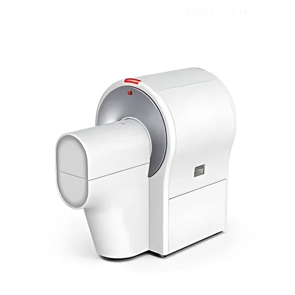
Aolong Micro CT100 Industrial Micro-CT System
| Brand | Aolong |
|---|---|
| Origin | Liaoning, China |
| Manufacturer Type | OEM Manufacturer |
| Country of Origin | China |
| Model | Micro CT100 |
| Price | Upon Request |
| X-ray Tube | 40–90 kV, 50 W, focal spot size 15 µm @ 6 W |
| Additional Filters | 6 metal filters (automated selection) |
| Detector | CMOS flat-panel detector, 1536 × 1944 pixels, 14-bit depth |
| Spatial Resolution | 2.0–44.9 µm (reconstruction-dependent) |
| Effective Field of View (FOV) | 80 × 200 mm |
| Voxel Size (Reconstruction) | down to 2 µm isotropic |
| Scan Time | 2 s (high-speed mode), 20 s (standard mode) |
| Dimensions | 880 × 1500 × 1500 mm (W × D × H) |
| Weight | < 950 kg |
| Radiation Safety | < 1 µSv/h at 10 cm from enclosure surface during operation |
Overview
The Aolong Micro CT100 is a high-performance, benchtop micro-focus X-ray computed tomography system engineered for non-destructive, three-dimensional structural and quantitative analysis across preclinical research, industrial materials inspection, and agricultural science. Based on cone-beam geometry and filtered back-projection reconstruction algorithms, the system utilizes a high-stability microfocus X-ray source (15 µm focal spot) and a large-area, low-noise CMOS flat-panel detector to deliver isotropic voxel resolution down to 2 µm. Its fully integrated hardware architecture—including motorized stage, automated filter wheel, thermally regulated animal handling platform, and lead-shielded enclosure—ensures compliance with international radiation safety standards (IEC 61331-1, IEC 62495) without requiring dedicated shielded rooms. Designed specifically for longitudinal in vivo imaging of small laboratory animals (mouse, rat) and ex vivo specimens, the Micro CT100 supports dynamic physiological monitoring via synchronized ECG, temperature, and respiratory gating interfaces—enabling functional morphometric assessment beyond static anatomy.
Key Features
- Sub-2 µm effective spatial resolution achieved through mechanical precision alignment, hardware-based geometric calibration, and iterative reconstruction optimization
- Automated sample recognition: intelligent carrier detection identifies specimen type (mouse/rat/ex vivo) and auto-configures optimal filter, FOV, kV, and exposure parameters
- Integrated multi-modal animal handling system: space-capsule-style carrier with built-in gas anesthesia delivery, waste anesthetic recovery, heated airflow control, ECG acquisition, thermal regulation, and escape-proof mechanical restraint
- Six-position automated metal filter wheel (Al, Cu, Sn, Mo, Ni, Pb-composite) minimizes beam hardening artifacts and reduces radiation dose by up to 40% compared to fixed-filter configurations
- Ultra-fast acquisition: 2-second high-speed scan mode enables rapid screening while maintaining sufficient SNR for bone mineral density (BMD) and trabecular architecture quantification
- Passive lead shielding embedded within the gantry housing ensures <1 µSv/h ambient dose rate at 10 cm—fully compliant with ALARA principles and local regulatory requirements for Class II B radiation devices
- Low-noise thermal management: proprietary fan array design maintains stable detector temperature and suppresses acoustic emissions below 45 dB(A)
Sample Compatibility & Compliance
The Micro CT100 accommodates live rodents (up to 500 g), excised tissues (e.g., femur, lung, heart, liver), polymer composites, ceramic scaffolds, seed samples, and electronic components. All scanning protocols adhere to ISO/IEC 17025 quality management requirements for measurement traceability. For preclinical applications, the system supports adherence to NIH Guide for the Care and Use of Laboratory Animals, AAALAC International standards, and FDA Guidance for Industry on Imaging Biomarkers (2021). DICOM 3.0 export ensures compatibility with PACS infrastructure and third-party analysis platforms. The integrated radiation safety interlock system complies with IEC 61000-6-4 (EMC) and meets CE marking requirements under the EU Medical Device Regulation (MDR 2017/745) Annex II classification for Class IIa in-vivo diagnostic imaging equipment.
Software & Data Management
Acquisition and reconstruction are managed via Aolong CT Suite v5.x—a validated, audit-trail-enabled software platform compliant with FDA 21 CFR Part 11 for electronic records and signatures. The suite includes GLP/GMP-ready modules for protocol versioning, user access control, instrument calibration logging, and raw projection data archiving. Quantitative analysis modules provide automated segmentation and morphometry for bone (BMD, BV/TV, Tb.Th, Tb.Sp, Tb.N), adipose tissue (subcutaneous, visceral, brown fat volume), lung parenchyma (airway lumen area, alveolar density), cardiac function (LV end-diastolic/systolic volume, ejection fraction), vasculature (vessel diameter, tortuosity, branching count), and organ volumetry (liver/kidney/spleen volume, surface area, sphericity). All results export to CSV, NIfTI, or STL formats; 3D visualization supports multi-planar reformatting, volume rendering, and distance/angle measurements with sub-voxel interpolation accuracy.
Applications
- Preclinical osteology: longitudinal monitoring of osteoporosis progression, fracture healing kinetics, osteoarthritis cartilage degradation, and biomaterial integration in orthopedic implants
- Metabolic phenotyping: volumetric quantification of adipose depot distribution and browning dynamics in diet-induced obesity and genetic models
- Pulmonary research: airway remodeling in asthma/COPD models, emphysema quantification, and inhalation drug deposition mapping
- Cardiovascular disease modeling: infarct sizing, myocardial wall thickening analysis, coronary calcification scoring, and aortic plaque characterization
- Vascular biology: microangiography of tumor neovasculature, cerebral microcirculation mapping, and stent deployment assessment
- Industrial QA/QC: porosity analysis in additive-manufactured metal parts, fiber orientation in composites, defect detection in castings, and grain structure visualization in alloys
- Agricultural science: seed viability assessment, root architecture phenotyping, and internal fruit defect detection without destructive sampling
FAQ
Is the Micro CT100 suitable for longitudinal in vivo studies?
Yes—the system’s low-dose acquisition modes, automated positioning repeatability (<5 µm), and physiological monitoring integration enable reliable serial imaging over weeks or months with minimal biological perturbation.
Does the software support automated bone morphometric analysis per ASBMR guidelines?
Yes—bone analysis modules implement standardized ASBMR nomenclature and calculation methods for trabecular and cortical parameters, including connectivity density, structure model index, and degree of anisotropy.
Can the system perform contrast-enhanced angiography?
Yes—when used with iodinated or nanoparticle-based contrast agents, the Micro CT100 achieves vessel contrast-to-noise ratios >15:1 at 10 µm resolution, supporting time-resolved perfusion studies.
What regulatory documentation is provided for GxP environments?
Aolong supplies IQ/OQ/PQ validation templates, software verification reports, radiation safety certificates, and a full 21 CFR Part 11 compliance package—including electronic signature implementation and audit trail configuration guidance.
Is remote operation supported?
Yes—secure TLS-encrypted remote desktop access and DICOM web viewer integration allow off-site protocol setup, scan monitoring, and preliminary image review without compromising data integrity or network security.


