Thermo Fisher EVOS XL / EVOS XL Core Cell Imaging System
| Brand | Thermo Fisher |
|---|---|
| Origin | USA |
| Manufacturer Authorization | Authorized Distributor |
| Import Status | Imported |
| Model | EVOS XL / EVOS XL Core |
| Pricing | Upon Request |
Overview
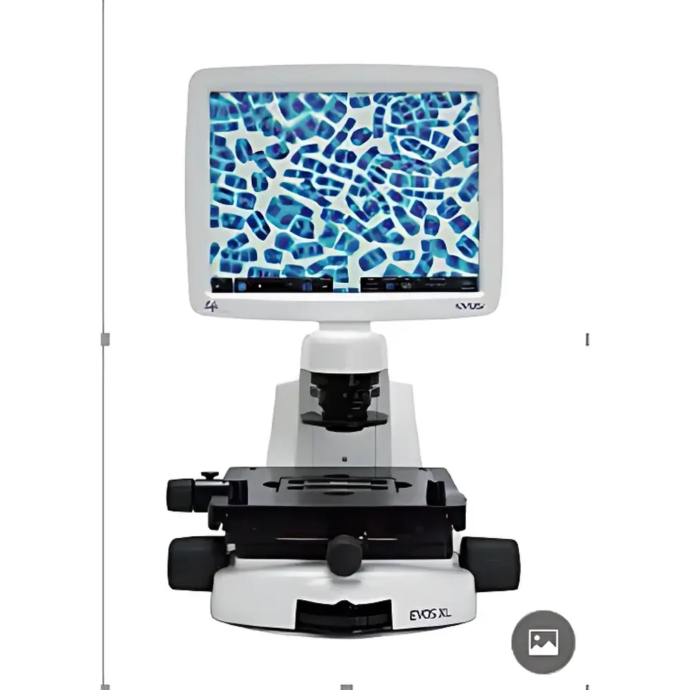
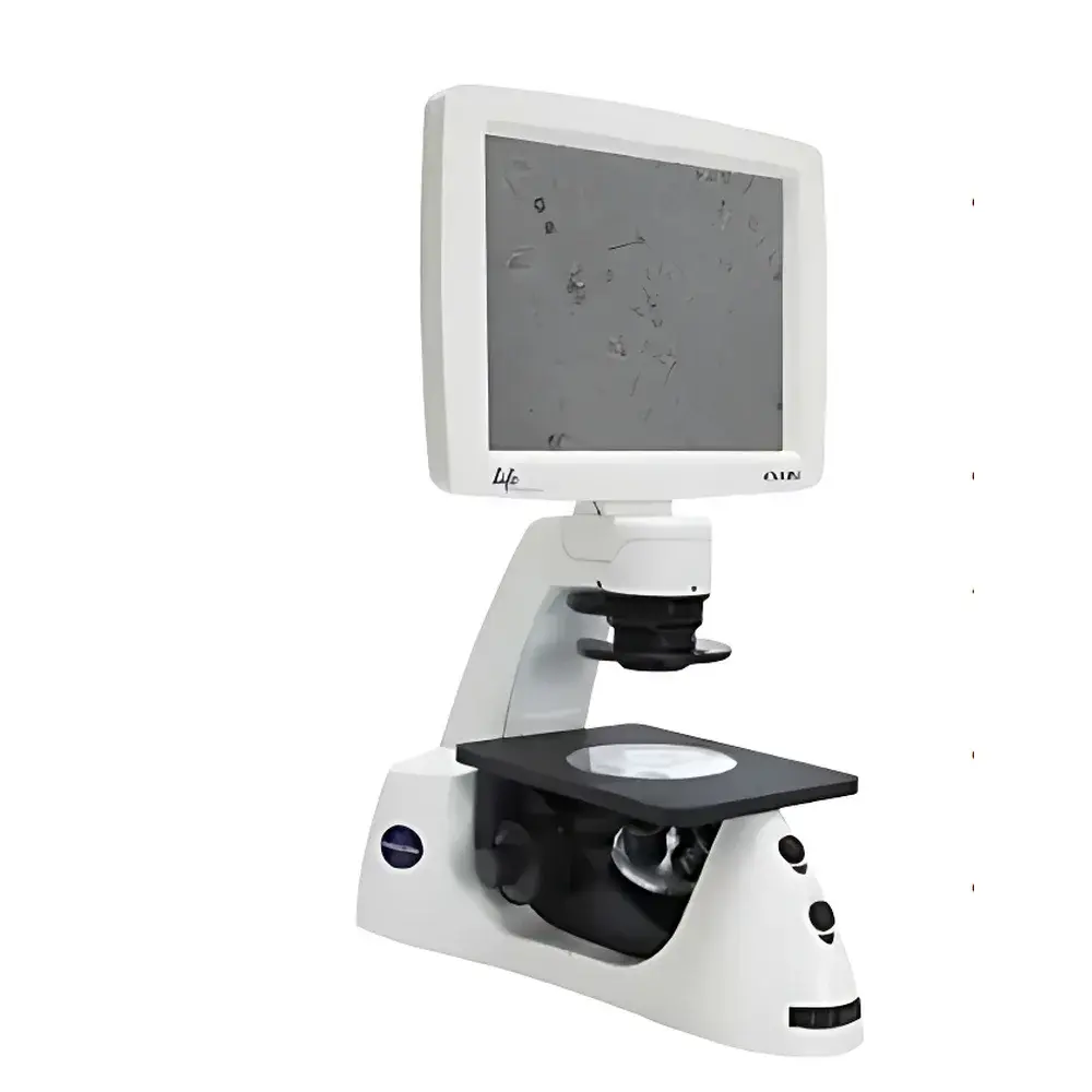
The Thermo Fisher EVOS XL and EVOS XL Core Cell Imaging Systems are benchtop, inverted digital microscopy platforms engineered for routine live-cell and fixed-sample visualization in bioscience laboratories. Unlike conventional compound microscopes requiring eyepieces and darkroom adaptation, the EVOS XL series integrates a high-resolution color CMOS sensor (24-bit/pixel), LED-based illumination, and an integrated 15.6-inch full-HD LCD display into a single compact unit—eliminating the need for external computers or ocular strain during extended observation sessions. The system operates on the principle of transmitted-light brightfield and phase contrast microscopy, enabling label-free, real-time assessment of cellular morphology, confluence, and tissue architecture without optical coupling complexity or alignment dependencies. Its modular optical path supports rapid interchange of objective lenses and contrast modules, delivering consistent image quality across diverse sample types while maintaining thermal and mechanical stability critical for longitudinal culture monitoring.
Key Features
- Integrated 15.6-inch HD LCD touchscreen display replaces traditional eyepieces—reducing visual fatigue and enabling ergonomic, seated operation.
- Dedicated hardware controls on the EVOS XL Core chassis: one-touch capture, onboard image storage (SD card slot), and intuitive navigation—optimized for high-throughput imaging workflows.
- Maintenance-free optical architecture: factory-aligned optics with no user-serviceable components; zero requirement for recalibration, collimation, or mechanical reassembly.
- Switchable contrast modes: seamless transition between brightfield and phase contrast via manual turret—no additional accessories or software toggling required.
- Long-life, stable-intensity LED illumination: rated for ≥50,000 hours with minimal spectral drift, ensuring reproducible exposure conditions across experiments and over time.
- Benchtop-compatible design: compact footprint and sealed internal electronics allow direct placement inside Class II biological safety cabinets or laminar flow hoods—enabling sterile imaging without sample transfer.
- 24-bit color depth CMOS imaging sensor: captures rich tonal gradation and true-color representation essential for H&E-stained histology sections, DAB immunohistochemistry, and unstained phase-dense structures such as mitotic spindles or nucleoli.
Sample Compatibility & Compliance
The EVOS XL platform accommodates standard tissue culture vessels—including 6–96-well plates, Petri dishes, chamber slides, and glass-bottom dishes—without stage modification. Its working distance and Z-axis clearance support both upright and inverted configurations for adherent and suspension cultures. While not intended for fluorescence applications (lacking excitation/emission filters or mercury/xenon sources), the system meets core requirements for GLP-compliant documentation in QC labs performing routine morphological assessments. Image metadata (timestamp, objective magnification, contrast mode, exposure settings) is embedded in EXIF headers and exportable in TIFF or JPEG formats compatible with LIMS integration. Though not FDA 21 CFR Part 11–validated out-of-the-box, audit trails and user-access logs can be implemented via networked file server protocols under institutional IT governance frameworks aligned with ISO/IEC 17025 and CLIA guidelines.
Software & Data Management
Imaging control and basic annotation are handled through the embedded EVOS Imaging Software v3.x, running natively on the device’s ARM-based OS. No PC dependency is required for acquisition, preview, measurement (e.g., confluence estimation, scale bar insertion), or export. Images are saved directly to internal flash memory or removable SD cards (up to 128 GB). Batch export supports DICOM-conformant naming conventions and folder structures suitable for PACS-adjacent archival. Raw image files retain full bit-depth fidelity and are compatible with third-party analysis tools including ImageJ/Fiji, MATLAB, and commercial packages like HALO or Visiopharm when exported in uncompressed TIFF format. Firmware updates are delivered via USB drive and include version-controlled release notes traceable to ISO 13485 manufacturing records.
Applications
- Routine monitoring of adherent mammalian cell lines (e.g., HEK293, HeLa, CHO) for passage timing, viability assessment, and contamination detection.
- Quantitative confluence analysis during transfection optimization, CRISPR editing validation, or bioprocess scaling studies.
- Stem cell culture management: tracking colony formation, differentiation progression (e.g., neural rosette emergence), and spontaneous beating in cardiomyocyte derivatives.
- Histopathology support: digitization of H&E- and DAB-stained paraffin sections for teaching archives, inter-laboratory comparison, or pre-screening prior to whole-slide scanning.
- Developmental biology: time-lapse documentation of embryoid body aggregation, gastruloid symmetry breaking, or organoid lumen formation—when paired with environmental chamber add-ons (sold separately).
- Quality control in biomanufacturing: verification of monolayer integrity post-thaw, scaffold seeding uniformity, or scaffold-free spheroid size distribution.
FAQ
Does the EVOS XL support fluorescence imaging?
No—the EVOS XL and XL Core models are optimized exclusively for transmitted-light brightfield and phase contrast. Fluorescence capability requires the separate EVOS FL Auto or EVOS M5000 platforms with dedicated light engines and filter cubes.
Can images be exported in scientific publication–ready formats?
Yes—TIFF exports preserve full 24-bit color depth and embedded scale information; JPEG outputs include adjustable compression levels. Both formats support inclusion of scale bars, annotations, and multi-panel figure assembly via bundled layout tools.
Is calibration required before first use?
No—optical alignment and illumination uniformity are factory-verified and locked. Users perform only a one-time screen brightness adjustment and objective identification during initial setup.
What is the maximum supported magnification?
With available objectives (4×, 10×, 20×, 40×), the system achieves up to 400× total magnification (40× objective × 10× digital zoom), though resolution remains constrained by the 1.3 NA limit of the air objectives and sensor pixel pitch.
How is data security managed in shared lab environments?
User accounts are not implemented at the device level; however, image files carry embedded timestamps and operator-assigned tags. For regulated environments, institutions typically enforce access control via network-level authentication and encrypted NAS storage policies compliant with HIPAA or GDPR.


