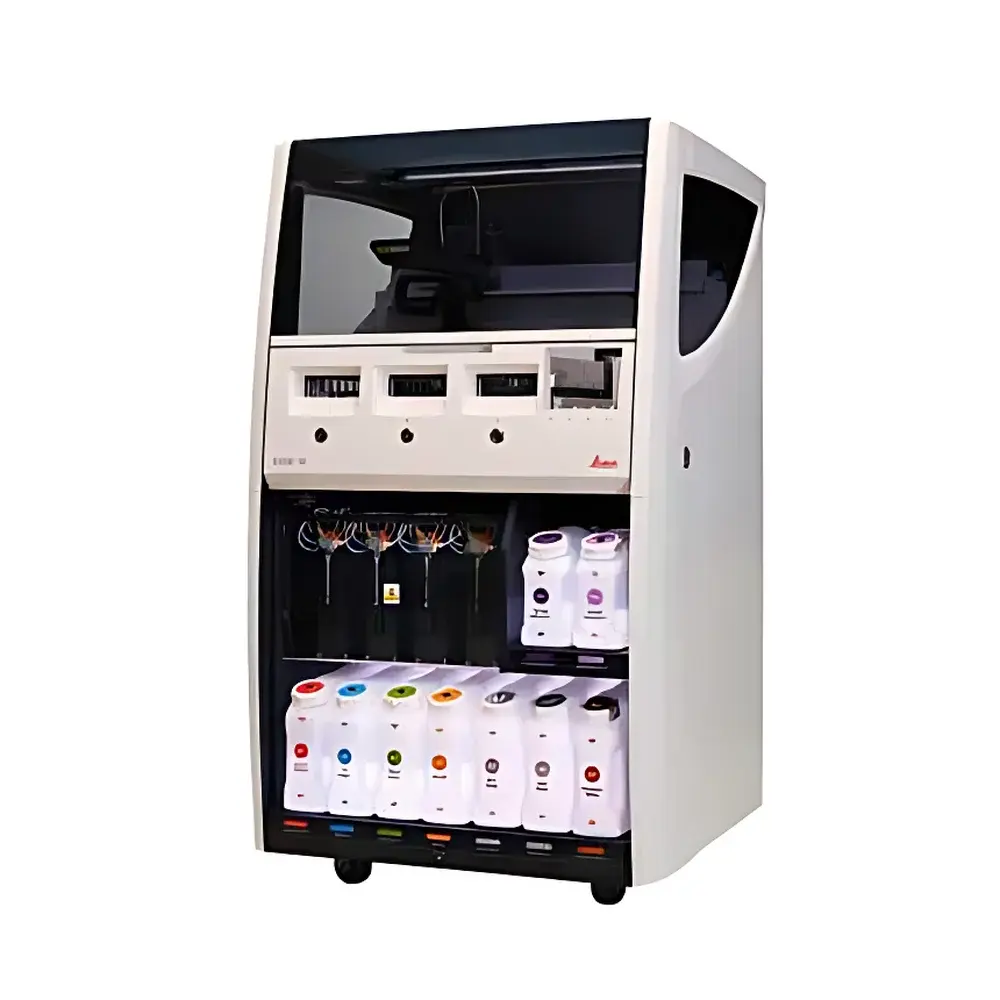
Leica Biosystems Bond-III Fully Automated Tissue Staining System
| Brand | Leica Biosystems |
|---|---|
| Origin | Germany |
| Model | Bond-III |
| Automation Level | Fully Automated |
| Staining Method | Immersion-Based |
| Max. Slides per Run | 30 |
| Max. Reagent Slots | 36 |
| Dimensions (W×H×D) | 790 × 1378 × 806 mm |
| Primary Applications | Immunohistochemistry (IHC), In Situ Hybridization (ISH) |
| Regulatory Compliance | CE-IVD, FDA-cleared for IVD use in the US |
| Software Platform | Bond RX™ Control Software v5.x |
| Connectivity | Ethernet, DICOM-SR export capable |
| Power Supply | 230 V AC, 50/60 Hz, 16 A |
Overview
The Leica Biosystems Bond-III is a fully automated, high-throughput tissue staining platform engineered for precision, reproducibility, and workflow integration in diagnostic and research pathology laboratories. Built upon Leica’s proven Bond technology architecture, the system employs immersion-based staining—where slides are submerged in reagent reservoirs under precisely controlled temperature, incubation time, and fluid exchange dynamics—to ensure uniform antigen retrieval, antibody binding, and detection chemistry across all tissue sections. Unlike air-gap or capillary-action systems, immersion delivery minimizes edge effects and enhances consistency in low-abundance target detection, particularly critical for multiplex IHC and dual ISH-IHC workflows. Designed for routine clinical diagnostics as well as translational research, the Bond-III supports end-to-end automation of antigen retrieval (heat-induced and enzymatic), primary/secondary antibody incubation, signal amplification, counterstaining, and dehydration—all within a single instrument footprint. Its modular reagent management and programmable protocol architecture enable seamless adaptation to evolving biomarker validation requirements and laboratory-specific SOPs.
Key Features
- Fully automated operation with walk-away capability: unattended processing of up to 30 slides per run, including deparaffinization, antigen retrieval, staining, and coverslipping-ready output.
- 36 independent reagent positions with integrated refrigerated storage (4–8 °C), enabling concurrent deployment of primary antibodies, polymer detection systems, chromogens, and nucleic acid probes without manual intervention.
- Precise thermal control across all modules: programmable water bath for antigen retrieval (up to 120 °C, ±0.5 °C stability), heated incubation chambers (30–45 °C, ±0.3 °C), and ambient-temperature reagent dispensing.
- Bond RX™ Control Software v5.x with intuitive drag-and-drop protocol builder, audit trail logging compliant with 21 CFR Part 11, and role-based user access controls for GLP/GMP environments.
- Integrated barcode scanning for slide and reagent tracking, supporting full traceability from specimen receipt through final stained slide generation.
- Compact footprint (790 × 1378 × 806 mm) optimized for space-constrained lab layouts while maintaining Class II biological safety compatibility when used with optional fume hood integration.
Sample Compatibility & Compliance
The Bond-III accommodates standard 1” × 3” glass microscope slides—including charged, APES-coated, and polymer-coated variants—with compatibility for both formalin-fixed paraffin-embedded (FFPE) and frozen tissue sections. It supports a broad range of tissue types (e.g., carcinoma, lymphoid, neural, and soft-tissue specimens) and thicknesses (3–5 µm). All protocols are validated against international standards including ISO 15189:2022 (medical laboratories), CAP Checklist ANP.42700 (IHC validation), and CLSI EP28-A3c (precision and linearity assessment). The system carries CE marking under IVDR (2017/746) and is FDA 510(k)-cleared for use with IVD-labeled assays in the United States. Reagent kits supplied by Leica Biosystems undergo rigorous lot-to-lot testing to ensure batch consistency and diagnostic reliability.
Software & Data Management
Bond RX™ Control Software provides centralized protocol management, real-time monitoring of run status, and comprehensive electronic recordkeeping. Each staining cycle generates a digitally signed audit log containing timestamps, operator ID, reagent lot numbers, temperature profiles, and fluid volume dispensed—fully compliant with FDA 21 CFR Part 11 requirements for electronic records and signatures. Data export options include CSV for LIMS integration and DICOM-SR for structured reporting in digital pathology workflows. Remote diagnostics and software updates are delivered via secure TLS-encrypted connection, with optional on-premise server deployment available for institutions requiring air-gapped network environments.
Applications
The Bond-III serves as a foundational platform for immunohistochemical (IHC) profiling in oncology diagnostics—including ER/PR/HER2 testing in breast cancer, PD-L1 expression scoring, and MSI/MMR marker panels—as well as RNA/DNA in situ hybridization (ISH) for ALK, ROS1, and NTRK gene fusions. Its flexibility extends to research applications such as spatial phenotyping, multiplexed biomarker co-localization studies, and pre-analytical optimization of novel antibody clones. Laboratories routinely deploy the system in high-volume surgical pathology, molecular diagnostics, and biobanking operations where reproducibility, throughput, and regulatory traceability are non-negotiable.
FAQ
What types of tissue sections can be processed on the Bond-III?
FFPE and frozen sections ranging from 3–5 µm thickness on standard glass slides; compatible with charged, APES-, and polymer-coated substrates.
Does the Bond-III support dual ISH-IHC staining on the same slide?
Yes—via sequential, programmable module scheduling that maintains optimal pH, temperature, and wash stringency between nucleic acid and protein epitope detection steps.
How is reagent consumption monitored and managed?
Each reagent slot includes integrated level sensors and barcode-scanned lot registration; software calculates remaining volume per protocol and triggers low-level alerts.
Can the Bond-III integrate with existing LIS or digital pathology platforms?
Yes—through HL7 v2.x messaging, DICOM-SR export, and configurable API endpoints for bidirectional data exchange.
Is on-site service and preventive maintenance available globally?
Leica Biosystems operates certified field service networks across 100+ countries, offering SLA-backed remote diagnostics, scheduled PM visits, and 24/7 technical support escalation paths.


