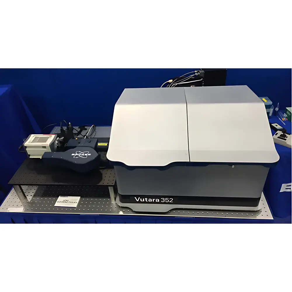
Bruker Vutura352 Dual-Focal-Plane Real-Time Super-Resolution Microscopy System
| Brand | Bruker |
|---|---|
| Origin | USA |
| Manufacturer Type | Authorized Distributor |
| Origin Category | Imported |
| Model | Vutura352 |
| Instrument Type | Optical Imaging System |
| Lateral Resolution | 20 nm |
| Axial Resolution | 50 nm |
| Maximum Frame Rate | 300 fps |
| Field of View | 40 µm × 40 µm |
| Sample Capacity | Single Specimen |
| Application Domain | Live-cell & Small-animal In Vivo Imaging |
Overview
The Bruker Vutura352 is a dual-focal-plane, real-time super-resolution microscopy system engineered for quantitative, three-dimensional single-molecule localization microscopy (SMLM) in living specimens. Unlike conventional widefield or confocal platforms, the Vutura352 employs a precisely calibrated dual-objective architecture to simultaneously acquire two distinct focal planes within a single exposure—enabling direct, model-free axial coordinate assignment without iterative fitting or point-spread-function (PSF) engineering. This optical design leverages astigmatic or biplane detection principles to resolve z-position with high fidelity, achieving an axial resolution of 50 nm and lateral resolution of 20 nm under physiological imaging conditions. The system is optimized for low-phototoxicity acquisition, supporting extended time-lapse SMLM of dynamic subcellular structures—including cytoskeletal remodeling, synaptic vesicle trafficking, and nuclear pore complex dynamics—in live cells and thin tissue preparations up to 15 µm depth. Its native compatibility with standard fluorescent probes (e.g., Alexa Fluor dyes, HaloTag ligands, and self-labeling proteins) ensures seamless integration into existing labeling workflows.
Key Features
- Dual-focal-plane simultaneous acquisition architecture for direct 3D single-molecule localization without computational z-reconstruction
- Real-time imaging capability up to 300 frames per second (fps), enabling capture of rapid molecular diffusion and transient protein–protein interactions
- Engineered optical path with minimal chromatic aberration and thermal drift compensation for high reproducibility across multi-hour acquisitions
- Integrated adaptive illumination control to balance signal-to-noise ratio and photobleaching kinetics during live-sample imaging
- Modular platform supporting co-registration with confocal scanning units for hybrid super-resolution + structural context imaging
- Automated focus stabilization via infrared-based objective position feedback, maintaining sub-10 nm axial stability over >8-hour sessions
Sample Compatibility & Compliance
The Vutura352 accommodates standard glass-bottom culture dishes (No. 1.5 coverslip), silicone chamber slides, and custom microfluidic devices compatible with inverted microscope configurations. It supports live mammalian cell lines (e.g., HeLa, U2OS, primary neurons), organotypic brain slices, and explanted tissues from murine models. All hardware and firmware comply with IEC 61000-6-3 (EMC emission standards) and IEC 60601-1 (medical electrical equipment safety). Data acquisition modules adhere to ALCOA+ principles for traceability and are configurable to meet GLP-compliant documentation requirements, including audit trails for acquisition parameters, timestamped metadata embedding, and user-access logging per FDA 21 CFR Part 11 guidelines.
Software & Data Management
Acquisition and reconstruction are managed through Bruker’s proprietary Vutura Acquisition Suite v4.x, which includes GPU-accelerated real-time localization engine (CUDA-enabled), drift correction via cross-correlation of fiducial beads or cellular landmarks, and batch-wise cluster analysis using DBSCAN or Ripley’s K-function. Export formats include TIFF (with OME-XML metadata), HDF5 (for large-scale time-series), and CSV-compatible localization tables compliant with the NanoJ-SQUIRREL and BigDataViewer interoperability standards. Raw data archives are structured according to the BIDS-Microscopy extension schema, facilitating FAIR data management and integration with institutional LIMS or ELN systems.
Applications
- Quantitative nanoscale mapping of membrane receptor clustering and nanodomain organization in live T-cells
- Dynamic tracking of RNA-binding protein condensates during stress granule assembly/disassembly
- In situ structural analysis of mitochondrial inner membrane cristae architecture in cardiomyocytes
- Long-term 3D SMLM of synapse formation and pruning in primary neuronal co-cultures
- Correlative imaging of tumor microvasculature using Vutura352 + Bruker’s Skyscan µCT for multi-scale structural–functional validation
FAQ
Does the Vutura352 support multi-color SMLM with standard organic dyes?
Yes—compatible with Alexa Fluor 488/568/647, CF dyes, and Janelia Fluor series; spectral separation achieved via dichroic filtering and sCMOS channel splitting.
Is axial calibration required before each experiment?
No—factory-calibrated dual-plane geometry is stored per objective lens; optional in-situ verification using 100-nm fluorescent beads is available via built-in calibration module.
Can the system be integrated into a cleanroom environment for sterile tissue imaging?
Yes—the optical head and sample stage are ISO Class 5 compatible; optional HEPA-filtered enclosure kits and glove-port interfaces are available as accessories.
What file formats are generated during acquisition and reconstruction?
Raw camera frames (.tiff), localized coordinates (.csv/.h5), rendered super-resolved images (.tiff with OME-XML), and acquisition logs (.json) are all natively exported.
Is remote operation supported for core facility deployment?
Yes—Vutura Acquisition Suite supports secure TLS-encrypted remote desktop access, role-based user permissions, and scheduled acquisition via REST API endpoints.


