Leica DM6 B Upright Intelligent Research Microscope
| Brand | Leica |
|---|---|
| Origin | Shanghai, China |
| Manufacturer Type | Authorized Distributor |
| Product Category | Domestic (China-assembled) |
| Model | DM6 B |
| Price Range | USD 55,000 – 68,000 (FOB Shanghai) |
| Microscope Type | Upright |
| Eyepiece Configuration | Binocular |
Overview
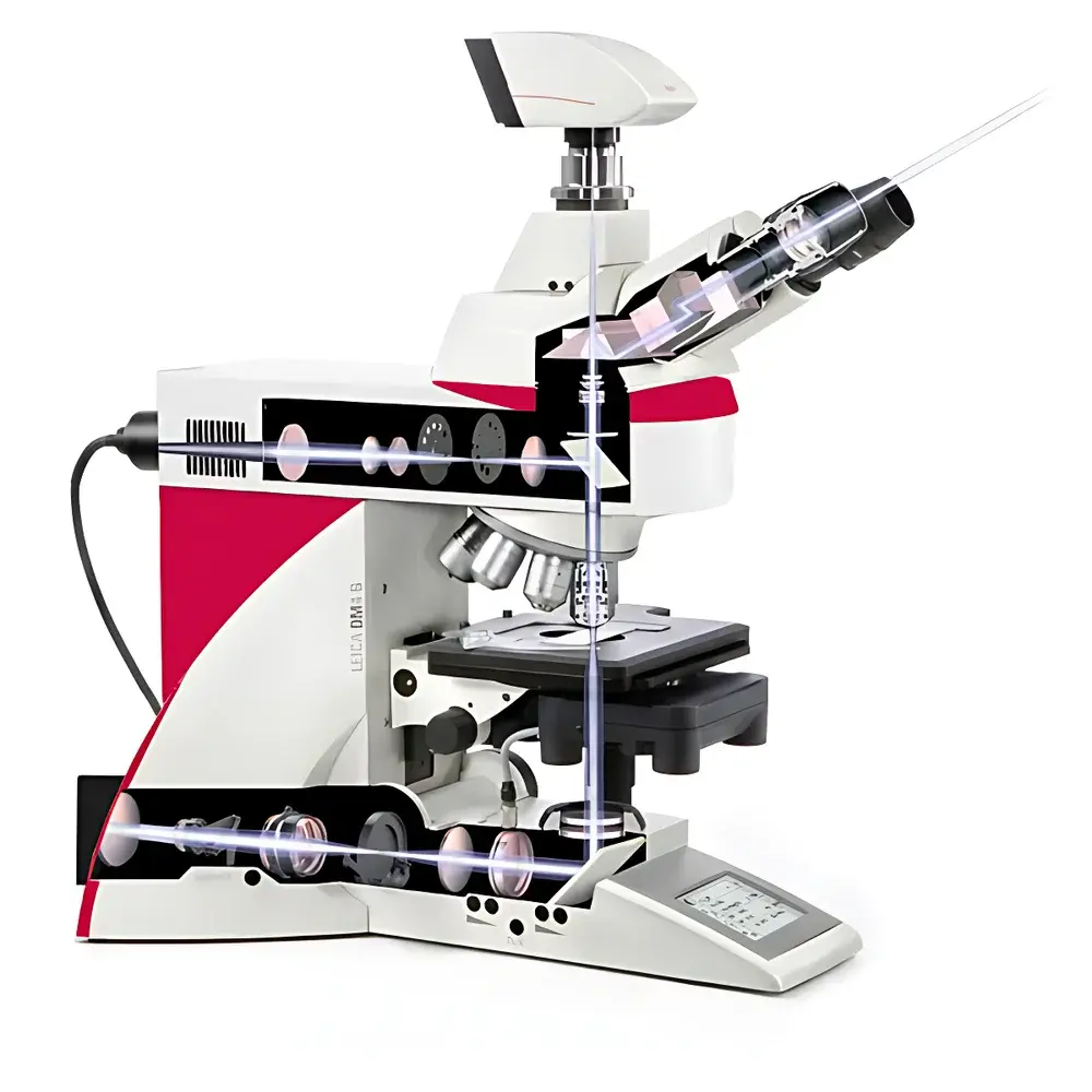
The Leica DM6 B is an upright intelligent research-grade microscope engineered for high-throughput life science laboratories, clinical pathology units, and academic core imaging facilities. Built upon Leica Microsystems’ decades of optical engineering heritage, the DM6 B implements a fully motorized, software-driven architecture that integrates hardware automation with intuitive digital workflow management. Its optical design follows Köhler illumination principles and supports multiple contrast modalities—including brightfield, phase contrast, differential interference contrast (DIC), and fluorescence—enabling rigorous morphological, cytological, and histological analysis. Unlike conventional manual microscopes, the DM6 B features embedded sensor feedback loops, programmable stage movement, and real-time objective lens recognition, ensuring repeatable, traceable, and audit-ready imaging sessions compliant with GLP and ISO/IEC 17025 laboratory practices.
Key Features
- Fully motorized upright platform with 7-position objective turret, encoded condenser, and automated focus drive for reproducible Z-stack acquisition
- 19-mm sCMOS-compatible imaging port optimized for standard scientific CMOS sensors (e.g., Hamamatsu ORCA-Fusion BT, PCO.edge 4.2), minimizing vignetting and maximizing signal-to-noise ratio
- Smart Illumination Management System supporting both halogen (3200 K) and LED (5700 K) light sources, with Constant Color Intensity Control (CCIC) ensuring stable white balance across intensity levels
- Integrated fluorescence excitation management (ExMan) and fluorescence intensity management (FIM) for precise spectral alignment and photobleaching mitigation
- Leica Application Suite X (LAS X) software with Smart Tile Scan, Virtual Slide Navigation, and Auto-Region-of-Interest (ROI) detection for rapid whole-slide surveying and targeted high-magnification imaging
- Over 300 Leica-certified objectives available—from 1.25× overview to 100× oil immersion—with chromatic aberration correction up to Plan Apochromat grade and transmission optimization for UV–NIR wavelengths
Sample Compatibility & Compliance
The DM6 B accommodates standard 1″ × 3″ glass slides, Petri dishes (up to 100 mm diameter), multi-well plates (6–96-well), and live-cell chambers with temperature/humidity control integration. Its modular stage design supports optional motorized XY translation (±50 mm travel), Z-height adjustment (0–30 mm clearance), and mechanical specimen holders compatible with cryo- or heated stages. The system complies with IEC 61000-6-3 (EMC emissions), IEC 61000-6-2 (immunity), and meets essential requirements of the EU Medical Device Regulation (MDR 2017/745) when configured for in vitro diagnostic applications. Software operation supports 21 CFR Part 11-compliant user authentication, electronic signatures, and full audit trail logging via LAS X Enterprise Edition.
Software & Data Management
Leica LAS X serves as the central interface for instrument control, image acquisition, annotation, measurement, and metadata tagging. It enables DIC-based quantitative phase imaging, fluorescence intensity profiling, and multi-channel spectral unmixing. All acquired images are stored with embedded EXIF-style metadata—including objective ID, exposure time, illumination intensity, filter set, and stage coordinates—ensuring FAIR (Findable, Accessible, Interoperable, Reusable) data handling. LAS X supports direct export to TIFF, OME-TIFF, and ND2 formats, and integrates with institutional LIMS and PACS systems via DICOM-SR and RESTful API. Batch processing workflows—including auto-focus calibration, brightness/contrast normalization, and ROI-based statistical reporting—are scriptable using Python-based LAS X Scripting Engine.
Applications
The DM6 B is routinely deployed in hematology for peripheral blood smear analysis; in histopathology for frozen section evaluation and tumor margin assessment; in stem cell research for colony morphology scoring and differentiation staging; and in neuroscience for Golgi-stained neuron tracing and immunofluorescent synaptic marker quantification. Its high-speed tile-scan capability supports digital pathology archival (e.g., Aperio-compatible SVS ingestion), while its low-phototoxicity LED illumination and ExMan module make it suitable for long-term live-cell time-lapse imaging under controlled environmental conditions. Academic labs use its modular configuration to support teaching modules on optical physics, contrast mechanisms, and quantitative microscopy validation.
FAQ
Is the DM6 B manufactured in Germany?
No—the DM6 B sold in mainland China is assembled at Leica’s Shanghai manufacturing facility under strict quality oversight from Wetzlar, Germany. Optical components (objectives, prisms, filters) are sourced from German production lines and undergo final calibration in Shanghai.
Does the system support third-party cameras?
Yes—any sCMOS or CCD camera with C-mount or F-mount interface and SDK support can be integrated via LAS X’s generic driver framework. Full hardware synchronization (trigger, exposure control) requires vendor-provided DLLs or GenICam compliance.
Can the DM6 B be used for routine clinical diagnostics?
Yes—when equipped with CE-IVD marked objectives, illumination modules, and LAS X Diagnostic Edition, the system meets EN ISO 15189 requirements for accredited medical laboratories.
What is the warranty and service coverage?
Standard coverage includes 24-month parts-and-labor warranty, remote diagnostics support, and annual preventive maintenance (PM) packages aligned with ISO/IEC 17025 calibration intervals.
How does CCIC differ from conventional halogen intensity control?
CCIC dynamically adjusts filament voltage and color-compensating filter position to maintain constant correlated color temperature (3200 K ±50 K) across 10–100% intensity range—eliminating white balance drift during multi-step imaging protocols.


