Leica EnFocus Intraoperative OCT Imaging System
| Brand | Leica |
|---|---|
| Country of Origin | Germany |
| Model | EnFocus |
| Regulatory Classification | Class II Medical Device (Imported) |
| Primary Application | Ophthalmic Surgery |
Overview
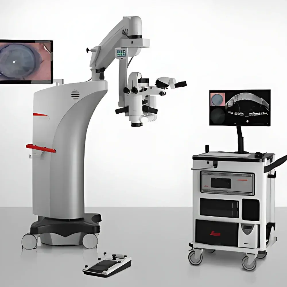
The Leica EnFocus Intraoperative OCT Imaging System is a high-precision, integrated optical coherence tomography (OCT) platform engineered for real-time, cross-sectional visualization of ocular microstructures during anterior and posterior segment surgery. Leveraging swept-source or spectral-domain OCT principles—depending on configuration—it delivers micron-level axial resolution and volumetric imaging capability directly within the surgical workflow. Unlike standalone clinical OCT systems, EnFocus is fully embedded into the Leica Proveo 8 ophthalmic microscope architecture, enabling seamless transition between conventional stereomicroscopy and depth-resolved OCT imaging without repositioning the patient or disrupting sterility. Its core function is to bridge the gap between preoperative diagnostic imaging and intraoperative tissue dynamics—providing objective, quantitative structural feedback at the point of intervention. This supports evidence-based decision-making in procedures where subtle anatomical shifts, fluid dynamics, or graft adherence cannot be reliably assessed by en face microscopy alone.
Key Features
- Fully integrated OCT module within the Leica Proveo 8 surgical microscope—no external scanners, cables, or secondary consoles required
- Sub-3 µm axial resolution (typical value: 2.4 µm in tissue) enabled by proprietary dispersion-compensated optics and high-sensitivity photodetectors
- Wide-field scanning capability: up to 20 × 20 mm field-of-view at high magnification—enabling full corneal coverage and peripheral monitoring without stage repositioning
- Real-time B-scan acquisition at up to 30 frames per second (fps), supporting dynamic assessment of tissue response during maneuvers such as DMEK graft unfolding or DALK dissection
- Four-quadrant display mode: simultaneous viewing of microscope view (top-left), en face view (bottom-left), and dual orthogonal OCT B-scans (right panel)
- Independent OCT control via footswitch, ergonomic handle, or 27″ HD touchscreen—designed for hands-free activation and parameter adjustment under sterile conditions
- Preset protocol management: customizable scan patterns, depth ranges, and measurement overlays assignable to hardware controls for procedure-specific workflows
- Integrated measurement suite: on-screen calipers for corneal thickness, graft-to-host alignment distance, needle insertion depth, and subretinal fluid height
Sample Compatibility & Compliance
The EnFocus system is validated for use with human ocular tissues in both anterior segment (e.g., cataract, keratoplasty, glaucoma drainage device placement) and select posterior segment applications (e.g., epiretinal membrane peeling, vitreoretinal interface assessment). It complies with IEC 60601-1 (medical electrical equipment safety), IEC 62304 (software lifecycle), and ISO 13485 (quality management systems). Regulatory clearance includes CE Marking (Class IIa), FDA 510(k) clearance (K171292), and China NMPA registration (National Medical Device Registration Certificate No. 20172162316). The system supports audit-ready data handling per FDA 21 CFR Part 11 requirements when paired with compatible DICOM-compliant recording solutions such as MedXchange Evolution4K. All image acquisition, annotation, and export functions maintain traceable user logs and timestamps for GLP/GCP-aligned documentation.
Software & Data Management
The EnFocus platform operates on the Leica InVivo software platform—a dedicated, intuitive interface optimized for intraoperative use. InVivo supports DICOM-compliant image capture, multi-frame playback (single-step or cine-loop), and side-by-side comparison of pre-/intra-/post-intervention scans. Measurement data—including layer thicknesses, distances, and angles—are exportable in CSV format for integration into electronic health records (EHR) or research databases. The system interfaces natively with Leica’s DI C800 digital eyepiece and offers four independent video outputs (HDMI/SDI) for OR-wide projection, PACS archiving, or simultaneous teaching display. Software updates follow a controlled release cycle with documented version history and validation reports available upon request for institutional IT compliance review.
Applications
- Anterior Segment Surgery: Real-time confirmation of Descemet’s membrane integrity during DMEK/DSAEK, precise depth control in DALK lamellar dissection, graft centration and apposition assessment in PKP or DALK, and intraoperative evaluation of glaucoma implant positioning
- Vitreoretinal Procedures: Identification of residual subretinal fluid post-vitrectomy, verification of internal limiting membrane peeling completeness, and detection of iatrogenic retinal breaks obscured by hemorrhage or vitreous haze
- Refractive & Corneal Surgery: Monitoring of femtosecond laser–created flap dimensions, stromal bed uniformity, and epithelial plug formation during SMILE or LASIK enhancement
- Surgical Training & Quality Assurance: Objective documentation of technical milestones, standardized assessment of trainee performance metrics, and retrospective analysis of procedural variability across cases
FAQ
Is EnFocus compatible with surgical microscopes other than Proveo 8?
No—EnFocus is a purpose-built, optomechanically integrated module designed exclusively for the Leica Proveo 8 platform. It is not retrofittable to prior-generation microscopes.
Does EnFocus require additional calibration before each procedure?
No routine recalibration is required between cases. The system performs automated optical alignment checks at startup and maintains calibration stability across temperature and humidity variations typical of modern OR environments.
Can OCT data be exported for offline analysis or publication?
Yes—raw and processed OCT volumes (B-scans, en face reconstructions, and measurement metadata) can be exported in standard formats (DICOM, TIFF, CSV) without proprietary codec restrictions.
What level of training is required for surgical staff?
Leica provides certified on-site clinical application specialist training covering system operation, interpretation fundamentals, workflow integration, and troubleshooting—typically completed in one full-day session per surgical team.
Is EnFocus approved for use in pediatric ophthalmic surgery?
Yes—clinical validation includes use in pediatric cataract and congenital glaucoma procedures; however, specific indications are subject to local regulatory labeling and institutional IRB protocols.


