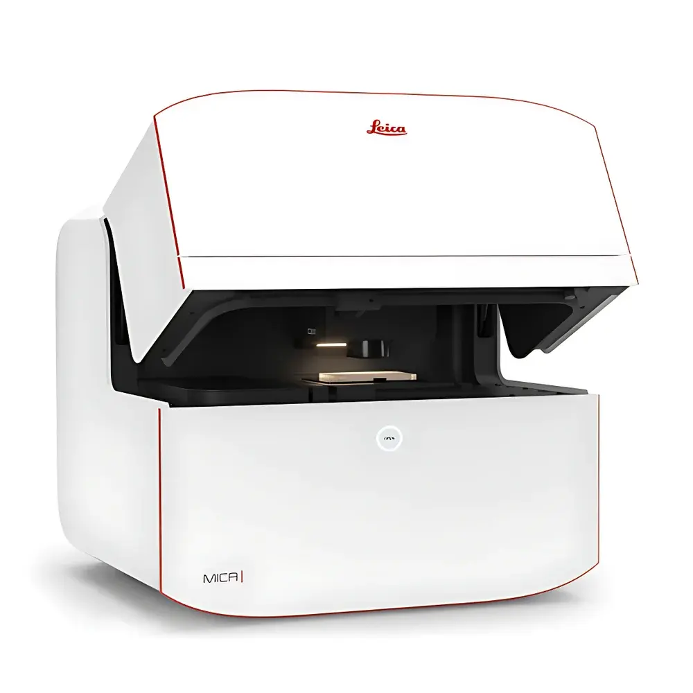
Leica MICA Multimodal Imaging and Analysis Platform
| Brand | Leica |
|---|---|
| Origin | Germany |
| Manufacturer Type | Authorized Distributor |
| Origin Category | Imported |
| Model | MICA |
| Price | USD 320,000 (FOB Hamburg) |
Overview
The Leica MICA Multimodal Imaging and Analysis Platform is an integrated, intelligent microscope system engineered for precision, reproducibility, and accessibility in both fixed and live-cell imaging workflows. Unlike conventional microscopes requiring sequential acquisition across multiple fluorescence channels or modality switching, MICA employs a unified optical architecture combining widefield and confocal imaging modalities within a single, fully motorized platform. Its core innovation lies in FluoSync — a proprietary spectral unmixing technology enabling simultaneous excitation and detection of up to four fluorescent labels without temporal or spatial misregistration. This ensures absolute spatiotemporal correlation across all channels, eliminating registration artifacts inherent in sequential scanning. The system operates on a principle of context-aware automation: hardware components (objectives, filter cubes, focus drives, light sources, detectors) are fully electrically controlled and coordinated through AI-guided software logic. Designed for compliance with GLP and GMP-aligned laboratory environments, MICA supports audit-trail-enabled operation per FDA 21 CFR Part 11 requirements when deployed with Leica LAS X Navigator and secure user authentication.
Key Features
- Fully automated multimodal imaging: seamless integration of widefield, confocal, phase contrast, and differential interference contrast (DIC) modes under one software-controlled workflow.
- FluoSync spectral decomposition: hardware-accelerated real-time unmixing of up to four fluorophores (e.g., DAPI, FITC, Cy3, Cy5) in a single exposure, preserving native emission spectra and eliminating crosstalk without linear unmixing assumptions.
- OneTouch adaptive illumination: intelligent selection of illumination intensity, exposure time, gain, and pinhole size based on user-defined priority — from “Sample Protection” (low-light, minimal phototoxicity) to “Image Quality” (maximized SNR).
- THUNDER computational clearing: GPU-accelerated deconvolution engine optimized for thick specimens; delivers optical sectioning quality comparable to confocal without mechanical pinhole constraints.
- LIGHTNING super-resolution enhancement: machine learning–based pixel-level detail recovery applied in real time during acquisition, compatible with both widefield and confocal data streams.
- Environmental chamber integration: temperature (20–40 °C ±0.2 °C), CO₂ (0–20% ±0.1%), and humidity (30–95% RH ±2%) control for long-term live-cell imaging over 72+ hours.
- Sample Finder autofocus: rapid generation of focus maps across large-area slides or multi-well plates using contrast-based Z-stack prediction and machine vision-assisted region-of-interest localization.
Sample Compatibility & Compliance
MICA accommodates standard microscopy formats including glass slides (1–3 mm thickness), 6–384-well plates, Petri dishes (35–100 mm), and chambered coverslips. It supports both fixed immunolabeled tissues (e.g., brain sections stained with DAPI, GFAP, NeuN, STL) and live 3D spheroids (MDCK, U2OS, primary neurons). All optical paths comply with ISO 10934-1:2005 (microscope nomenclature) and ISO 9022-3:2015 (environmental testing for optical instruments). For regulated environments, optional validation packages support IQ/OQ/PQ documentation aligned with ASTM E2500-13 and EU Annex 11 guidelines. Data integrity is maintained via encrypted local storage, timestamped metadata embedding (EXIF + custom XML), and role-based access control.
Software & Data Management
Leica LAS X Navigator serves as the central interface, supporting drag-and-drop experimental design, protocol templating, and version-controlled parameter inheritance across users and projects. AI-powered Pixel Classifier enables supervised training directly on acquired images using intuitive polygon and brush annotation tools — no coding required. Trained models are exportable as .lpx files and reusable across instrument deployments. All image processing steps (THUNDER, LIGHTNING, FluoSync unmixing) are non-destructive and logged with full provenance. Raw data is stored in standardized OME-TIFF format compliant with Bio-Formats and OMERO ingestion pipelines. Optional integration with Leica Application Suite X Cloud enables remote monitoring, collaborative annotation, and FAIR-compliant metadata tagging (MIAME/MINSEQE).
Applications
- High-content screening: Simultaneous 4-channel acquisition in multiwell plates reduces cycle time by ≥4× versus sequential confocal scanning; ideal for caspase-3/7 apoptosis assays using CellEvent™, TMRE, SiR-Actin, and DAPI.
- 3D tissue analysis: Correlative widefield overview (20×) and confocal zoom (63× water immersion, NA 1.20) enable hierarchical interrogation — from crypt architecture in colon sections to subcellular tubulin detyrosination patterns.
- Long-term live-cell dynamics: Stable environmental control permits >72 h timelapse of GFP-tagged MX1 spheroids at 30 min intervals, with automatic focus drift correction and phototoxicity minimization via OneTouch Sample Protection mode.
- Mitochondrial functional imaging: Dual-probe assessment (MitoTracker Green + TMRE) with FluoSync unmixing yields quantitative metrics on mass vs. membrane potential without channel bleed-through or temporal offset.
- Neuroscience research: Co-registration of neuronal nuclei (DAPI), astrocytes (GFAP-Cy3), synaptic terminals (STL-FITC), and newborn neurons (NeuN-Cy5) in rat brain sections at 10× tiling resolution, achieving full spatial fidelity across 100+ fields of view.
FAQ
Does MICA require external computing hardware for FluoSync or THUNDER processing?
No — all real-time spectral unmixing and computational clearing are executed on-board using dedicated FPGA and NVIDIA GPU modules integrated into the main controller unit.
Can MICA be used for super-resolution applications beyond LIGHTNING?
LIGHTNING is not true super-resolution (e.g., STED or PALM); it enhances effective resolution through learned priors. For diffraction-unlimited techniques, Leica recommends pairing MICA with STED systems via shared sample stages and synchronized acquisition protocols.
Is MICA validated for use in clinical diagnostics laboratories?
While MICA meets IEC 61010-1 safety standards and supports 21 CFR Part 11 compliance, its use in diagnostic reporting requires site-specific analytical validation per CLIA/CAP or ISO 15189 requirements — not provided out-of-the-box.
How does MICA handle refractive index mismatches in thick samples?
The system includes motorized correction collar objectives (e.g., 63×/1.20 CS2 Water MotCORR) and software-driven spherical aberration compensation algorithms calibrated per immersion medium and sample depth.
What file formats are supported for export and third-party analysis?
Native export includes OME-TIFF (with full metadata), TIFF stacks, and HDF5. Batch conversion to NRRD, CZI, or ND2 is supported via Leica’s command-line converter toolset compatible with Python, MATLAB, and Fiji/ImageJ scripting environments.


