Xunshu MCN Automated Micronucleus Analysis System for Erythrocytes
| Brand | Xunshu |
|---|---|
| Origin | Zhejiang, China |
| Manufacturer Type | Direct Manufacturer |
| Region of Origin | Domestic (China) |
| Model | MCN |
| Pricing | Available Upon Request |
Overview
The Xunshu MCN Automated Micronucleus Analysis System is a dedicated digital cytogenetic platform engineered for high-throughput, standardized micronucleus (MN) assay execution in mammalian erythrocytes—specifically polychromatic erythrocytes (PCEs) from bone marrow or peripheral blood. It implements a validated, image-based workflow compliant with OECD Test Guideline 474 and ISO 10993-3 for genotoxicity assessment. The system operates on the principle of quantitative morphometric analysis of Giemsa-stained cytological preparations, where micronuclei are identified as small, round, chromatin-positive structures within PCEs—distinct from main nuclei by size (1/20–1/5 the diameter of the PCE nucleus), staining intensity, and spatial separation. Unlike conventional manual scoring or threshold-based segmentation tools, the MCN system integrates adaptive stochastic resonance (ASR) signal enhancement to recover low-contrast PCE features obscured by heterogeneous background staining or suboptimal chromatin density—thereby improving detection sensitivity without compromising specificity.
Key Features
- Adaptive Stochastic Resonance Engine: Enhances weak-color signal-to-noise ratio in Giemsa-stained PCEs by leveraging nonlinear bistable system dynamics and mutual information entropy optimization—enabling robust segmentation of pale-staining PCEs against variable background intensities.
- Deep Morphological Learning Architecture: Trained on curated reference datasets of rodent and human erythrocyte morphology; supports automated discrimination between normochromatic erythrocytes (NCEs), PCEs, lymphocytes, granulocytes, and platelet fragments using multi-feature decision trees (nuclear texture, cytoplasmic hue saturation, contour convexity, and size distribution).
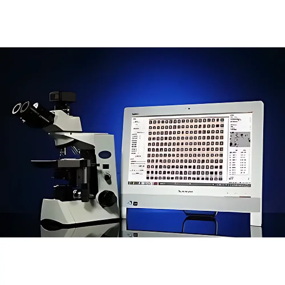
- High-Fidelity Image Acquisition Subsystem: Equipped with Plan-Apochromat oil-immersion objectives (100×, NA ≥ 1.4), large-format monochrome CCD sensor (≥ 5.0 MP, 12-bit dynamic range), and precision motorized stage with sub-micron positioning repeatability—ensuring consistent focus, illumination uniformity, and cellular detail preservation across fields of view.
- Automated MN Scoring Pipeline: Processes ≥2,000 PCEs per slide in <60 seconds; computes %MN-PCE (micronucleated PCEs / total PCEs × 100), %PCE/(PCE+NCE), and raw counts with audit-trail logging per field and cell.
- Interactive Verification Interface: All detected PCEs, NCEs, and MN-positive cells are stored with full metadata—including original coordinate position, acquisition timestamp, focal plane, and raw pixel data—allowing traceable re-evaluation, manual override, and annotation export in TIFF/PNG/CSV formats.
Sample Compatibility & Compliance
The MCN system is validated for use with Giemsa-stained smears prepared from murine, rat, and human bone marrow aspirates or peripheral blood samples processed according to standard protocols (e.g., ICH S2(R2), OECD TG 474). It accommodates standard 26 × 76 mm glass slides with coverslips (0.13–0.17 mm thickness). Data output complies with GLP documentation requirements: each analysis session generates an immutable PDF report containing instrument ID, operator ID, calibration timestamps, image acquisition parameters, cell count histograms, and statistical summaries. Audit trail functionality meets FDA 21 CFR Part 11 readiness criteria for electronic records and signatures when deployed with validated network storage and user access controls.
Software & Data Management
The proprietary Xunshu ImageStudio v5.x software provides integrated acquisition, processing, quantification, and reporting modules. All image operations—including deconvolution-enhanced sharpening, adaptive background flattening, morphological filtering, edge detection (Canny/Sobel), and non-linear contrast stretching—are implemented in GPU-accelerated C++ libraries. Measurement tools include calibrated digital micrometry (linear, angular, area, arc-length), particle counting with customizable ROI masking, and comet assay parameter extraction (tail moment, Olive tail moment, %DNA in tail). Export options support FAIR data principles: CSV for statistical analysis (R, Python, GraphPad), TIFF/OME-TIFF for archival, and DICOM-SR for PACS integration in translational research environments.
Applications
- Regulatory genotoxicity screening in pharmaceutical preclinical development (ICH M7, S2(R2))
- Environmental mutagen monitoring (e.g., wastewater effluents, airborne particulates)
- Occupational health surveillance programs for chemical exposure assessment
- Academic research in DNA repair mechanisms, chromosomal instability, and clastogenic/aneugenic agent profiling
- Quality control of biologics and cell therapies where genomic integrity is critical
FAQ
Does the MCN system support batch processing of multiple slides without manual intervention?
Yes—the motorized slide loader accepts up to 50 standard slides; fully automated scanning, focusing, and analysis are initiated via scheduled job queues.
Can the system be validated for GMP-compliant laboratories?
Yes—IQ/OQ/PQ documentation templates and 21 CFR Part 11 configuration guidance are provided; validation support includes installation qualification checklists and performance verification protocols using NIST-traceable microsphere standards.
Is training and technical support available internationally?
Yes—remote installation assistance, online SOP workshops, and annual software maintenance with version-controlled updates are included in extended service contracts.
What file formats are supported for raw image export and third-party analysis?
Raw 12-bit TIFF, OME-TIFF with embedded metadata, and lossless PNG; measurement data exports to CSV, Excel (.xlsx), and JSON for integration with LIMS or ELN platforms.
How does the system handle variability in Giemsa staining intensity across batches?
The ASR engine dynamically adjusts signal amplification thresholds per field-of-view based on local histogram statistics—eliminating need for manual color balance or global threshold tuning.
Related Products


