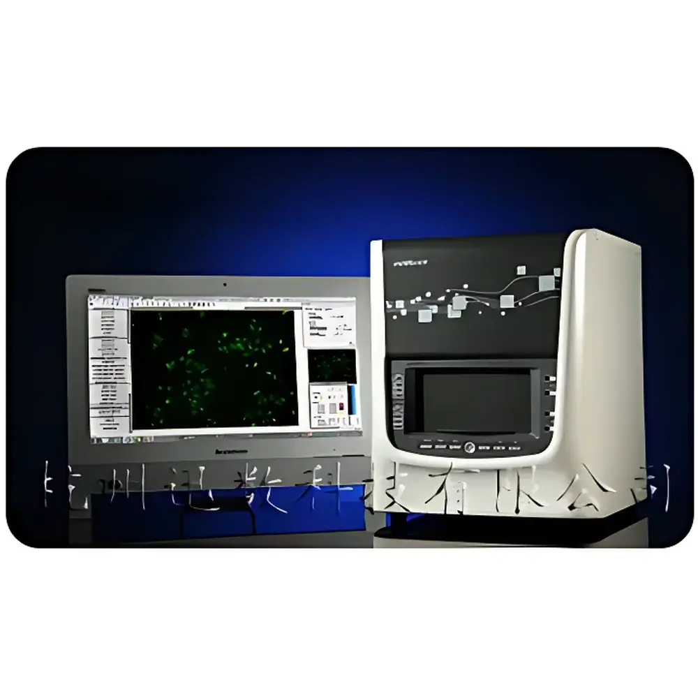
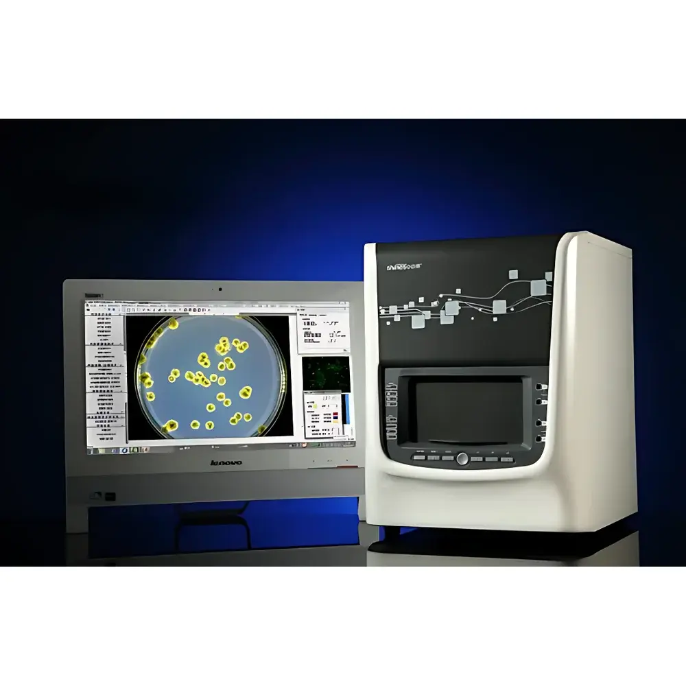
Xunshu SupcreG1 All-in-One Colony Counter, Antibiotic Zone Analyzer & Strain Screening System
| Brand | Xunshu |
|---|---|
| Origin | Zhejiang, China |
| Manufacturer Type | OEM Manufacturer |
| Region of Origin | Domestic (China) |
| Model | SupcreG1 |
| Price Range | USD 7,000–11,500 |
| Instrument Type | Fully Automated Colony Counter |
| Counting Speed | <1 second for up to 500 colonies |
Overview
The Xunshu SupcreG1 is a research-grade, integrated microbiological imaging and analysis platform engineered for precision colony enumeration, antibiotic inhibition zone measurement, and strain-level phenotypic screening. Unlike conventional colony counters limited to binary detection, the SupcreG1 employs a multi-modal optical architecture—combining tunable trichromatic LED illumination, dual-wavelength UV excitation (254 nm and 366 nm), and dual-path (epi- and trans-illumination) light delivery—to resolve morphological, chromatic, and fluorescent features across diverse microbial specimens. Its core analytical engine implements fourth-generation image segmentation algorithms based on level-set active contour models—including RGB-constrained color segmentation, multiphase level-set decomposition, and fast adaptive contour evolution—enabling robust separation of overlapping, irregular, or low-contrast colonies from heterogeneous agar backgrounds. Designed for GLP-compliant laboratories, the system supports traceable, auditable workflows aligned with ISO 21528-2, ISO 4833-1, and USP / microbiological testing standards.
Key Features
- Full-spectrum optical imaging: Adjustable correlated color temperature (3500K–8500K) trichromatic LED ring light ensures biologically faithful color reproduction—critical for chromogenic medium interpretation and strain differentiation.
- Dual-path illumination system: Top-mounted 360° flexible diffuse lighting enhances surface texture (e.g., rugosity, margin serration); bottom-mounted crystal-clear dark-field illumination resolves hyphal architecture in molds and actinomycetes—including intramycelial vs. aerial mycelium distinction.
- Integrated UV functionality: 254 nm lamp enables in-chamber sterilization and UV mutagenesis; dual-side 366 nm illumination facilitates fluorescence visualization of E. coli, coliforms, and GFP-expressing strains without external excitation sources.
- Enclosed dark-box imaging chamber: Human-centered hinged viewport door eliminates ambient stray light, suppressing refraction artifacts (e.g., halos, glare) at Petri dish interfaces—essential for high-fidelity colony boundary detection.
- Advanced segmentation algorithms: Proprietary “RGB-constrained level-set clustering” enables simultaneous multi-color colony classification on chromogenic media; “fast active contour” model delivers sub-pixel edge accuracy even on confluent or filamentous growths.
Sample Compatibility & Compliance
The SupcreG1 accommodates standard and specialized microbiological formats: pour plates, spread plates, membrane filters (including black-grid variants), spiral plates, multi-well plates (6–96-well), and 3M™ Petrifilm™ assays. It complies with regulatory imaging requirements for pharmaceutical QC (FDA 21 CFR Part 11-ready audit trail), food safety (ISO 7218, ISO 6887-1), and clinical microbiology (CLSI M07-A11). Built-in calibration protocols support traceable metric conversion (e.g., CFU/mL) using user-defined plate diameter and dilution factors. All image metadata—including illumination settings, algorithm parameters, operator ID, and timestamp—are embedded and exportable for full data integrity.
Software & Data Management
The bundled analysis software provides role-based access control, electronic signature capability, and configurable audit logging per FDA 21 CFR Part 11 and EU Annex 11. Statistical outputs—including total counts, size-classified distributions, zone diameters, area ratios (e.g., inhibition zone / colony diameter), and morphometric descriptors (area, perimeter, major/minor axis, circularity)—are exportable to Excel with raw image links. The database supports structured queries by sample ID, date range, assay type, or operator. Image processing tools include grayscale inversion, contrast/RGB channel adjustment, adaptive sharpening, grid background removal, and manual contamination masking—all non-destructive and versioned.
Applications
- Antibiotic susceptibility testing: Szone multi-mode zone analysis (auto-edge detection, circle-fitting approximation, 3-point manual definition) quantifies inhibition zones per CLSI guidelines.
- Enzyme or metabolite screening: Dual-ring analysis automatically computes diameter/area ratios for transparent zones (e.g., amylase, lipase), color-change zones (e.g., pH indicators), or growth-promotion zones (e.g., siderophore assays).
- Mold and filamentous organism quantification: “Mold One-Touch Measurement” extracts area, perimeter, Feret diameter, and aspect ratio from diffuse colonies via single-point edge initialization—eliminating subjective cross-diameter estimation.
- Vaccine and serology validation: Multi-region parallel counting supports OPKA (opsonophagocytic killing assay) and SBA (serum bactericidal assay) plate layouts with independent region masking and statistical aggregation.
- Fluorescent strain isolation: 366 nm excitation enables rapid visual identification and spatial mapping of GFP-tagged or auto-fluorescent isolates prior to picking.
FAQ
Does the SupcreG1 support regulatory compliance for pharmaceutical microbiology?
Yes—it meets data integrity requirements of FDA 21 CFR Part 11 and EU GMP Annex 11 through electronic signatures, immutable audit trails, and role-based permission controls.
Can it distinguish between different bacterial species on chromogenic media?
It performs automated multi-color clustering using RGB-constrained level-set segmentation, enabling simultaneous classification and enumeration of up to six color-defined populations per plate.
Is calibration traceable to international standards?
All geometric measurements are calibrated using NIST-traceable reference disks; software includes both automatic and manual recalibration routines with deviation logging.
What types of UV applications does the dual-wavelength system support?
254 nm enables routine chamber decontamination and targeted UV mutagenesis; 366 nm excites endogenous fluorophores (e.g., pyoverdine) and exogenous tags (e.g., GFP, RFP) without external filter cubes.
How does the system handle overlapping or chain-forming colonies?
Its “adaptive contour splitting” algorithm uses local intensity gradient analysis to separate adherent or filamentous colonies—configurable by user-defined separation thresholds and morphological constraints.


