NIUMAG NM42-060H Preclinical MRI System for Spontaneous Tumor Monitoring in Aging Mice
| Brand | NIUMAG |
|---|---|
| Origin | Jiangsu, China |
| Manufacturer Type | Authorized Distributor |
| Country of Origin | China |
| Model | NM42-060H |
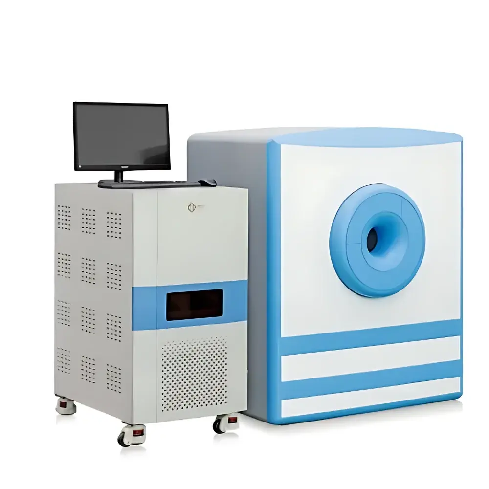
| Instrument Type | Preclinical Magnetic Resonance Imaging (MRI) System |
| Static Field Strength | 1.0 ± 0.05 T |
| Larmor Frequency | ~42 MHz |
| Bore Diameter | 60 mm |
| Application Scope | In vivo MRI of mice and rats using MR contrast agents |
| Target Applications | Spontaneous tumor detection (brain, subcutaneous, liver), longitudinal tumor growth & therapy response monitoring, obesity phenotyping, MR contrast agent development |
Overview
The NIUMAG NM42-060H is a dedicated benchtop preclinical magnetic resonance imaging (MRI) system engineered for non-invasive, longitudinal monitoring of spontaneous tumor development in aging murine models. Operating at a static magnetic field strength of 1.0 ± 0.05 T—corresponding to a proton Larmor frequency of approximately 42 MHz—the system delivers high signal-to-noise ratio (SNR) imaging optimized for small-animal physiology. Unlike terminal histopathological endpoints, this MRI platform enables repeated, quantitative volumetric assessment of tumor burden, spatial distribution, and microstructural evolution over time, preserving animal viability and experimental continuity. Its 60 mm horizontal bore accommodates standard rodent physiological monitoring accessories (respiratory gating, temperature control, ECG leads), ensuring stable acquisition conditions during multi-session studies. The system adheres to fundamental principles of spin echo and gradient echo MRI physics, supporting T1-, T2-, and proton density-weighted contrast generation—critical for distinguishing tumor heterogeneity, necrosis, edema, and contrast enhancement kinetics.
Key Features
- Dedicated 1.0 T permanent magnet architecture with active shimming for field homogeneity < 10 ppm over a 30 mm DSV (Diameter Spherical Volume)
- Integrated 60 mm inner-diameter RF transmit/receive coil optimized for mouse and rat torso/brain imaging
- Software-controlled pulse sequence library including spin echo (SE), fast spin echo (FSE), gradient echo (GRE), and inversion recovery (IR) protocols
- Real-time image reconstruction engine enabling on-the-fly preview of axial, coronal, and sagittal slices
- Physiological monitoring interface compatible with commercial small-animal anesthesia and vital sign systems
- Modular design supporting future upgrades to diffusion-weighted imaging (DWI) and dynamic contrast-enhanced (DCE-MRI) capabilities
Sample Compatibility & Compliance
The NM42-060H supports live imaging of C57BL/6, BALB/c, FVB/N, and other commonly used murine strains aged 18–30 months—models exhibiting high incidence of spontaneous mammary adenocarcinoma, lymphoma, and hepatocellular carcinoma. It is fully compatible with FDA-approved and research-grade MR contrast agents (e.g., Gd-DTPA, iron oxide nanoparticles) for enhanced lesion delineation and vascular permeability quantification. All imaging protocols comply with ARRIVE 2.0 guidelines for reporting animal research and support adherence to institutional IACUC requirements. Data output formats (DICOM 3.0, NIfTI) ensure interoperability with third-party analysis platforms such as ITK-SNAP, FSL, and MATLAB-based toolboxes used in academic and pharmaceutical preclinical labs.
Software & Data Management
The system runs NIUMAG’s proprietary AcqView™ acquisition software and AnalyzePro™ post-processing suite. AcqView™ provides intuitive sequence parameter tuning—including TR/TE/TI selection, matrix size (up to 512 × 512), slice thickness (0.3–2.0 mm), and number of averages—with built-in quality assurance checks prior to scan initiation. AnalyzePro™ delivers semi-automated tumor segmentation via region-growing and threshold-based algorithms, generating volumetric metrics (mm³), mean signal intensity, and normalized enhancement ratios. Audit trails log all user actions, parameter modifications, and calibration events—meeting GLP-aligned documentation standards. Export options include CSV for statistical packages (Prism, R), TIFF/PNG for publication-ready figures, and DICOM archives compliant with PACS integration in multi-site consortia.
Applications
- Longitudinal tracking of spontaneous tumor onset, growth kinetics, and metastatic spread in immunocompetent aging mice without euthanasia-induced endpoint bias
- Quantitative evaluation of therapeutic efficacy across modalities: chemotherapy, radiotherapy, immune checkpoint inhibitors, and targeted kinase inhibitors
- Correlative imaging for validation of novel PET/MRI radiotracers or nanoparticle-based contrast agents
- Phenotypic characterization of metabolic comorbidities—including hepatic steatosis and adipose tissue distribution—in tumor-bearing cohorts
- Preclinical biomarker discovery through texture analysis (radiomics) of T2-weighted and DCE-MRI datasets
- Support for IND-enabling toxicology and pharmacokinetic/pharmacodynamic (PK/PD) studies under GLP-like operational frameworks
FAQ
What is the minimum detectable tumor volume with the NM42-060H?
Detection sensitivity depends on contrast mechanism, acquisition parameters, and anatomical location; typical in vivo resolution supports reliable identification of lesions ≥1.5 mm³ in subcutaneous and hepatic sites under optimized T2-FSE protocols.
Can the system be used for functional MRI (fMRI) or diffusion imaging?
Standard configuration supports structural MRI only; optional diffusion gradient hardware and EPI-capable sequences are available upon request for DWI and ADC mapping.
Is the system compatible with stereotactic surgery platforms?
Yes—the 60 mm bore and modular coil design allow integration with commercially available murine stereotactic frames for guided intracranial tumor implantation followed by serial imaging.
Does the software support automated registration across longitudinal sessions?
AnalyzePro™ includes rigid and affine co-registration tools aligned to a standardized mouse brain atlas (DSURQE) or custom reference volumes, enabling voxel-wise change analysis over time.
What regulatory documentation is provided for audit readiness?
NIUMAG supplies IQ/OQ documentation templates, system calibration logs, DICOM conformance statements, and traceable field homogeneity reports—supporting internal QA and external regulatory review per ISO 13485 and FDA guidance for preclinical imaging devices.


